. 2020 Jan;17(1):85-94.
doi: 10.1038/s41423-018-0195-8. Epub 2019 Jan 11.

Intracellular HSP70L1 inhibits human dendritic cell maturation by promoting suppressive H3K27me3 and H2AK119Ub1 histone modifications

Affiliations

Intracellular HSP70L1 inhibits human dendritic cell maturation by promoting suppressive H3K27me3 and H2AK119Ub1 histone modifications

Lin Yi et al. Cell Mol Immunol. 2020 Jan.

Abstract

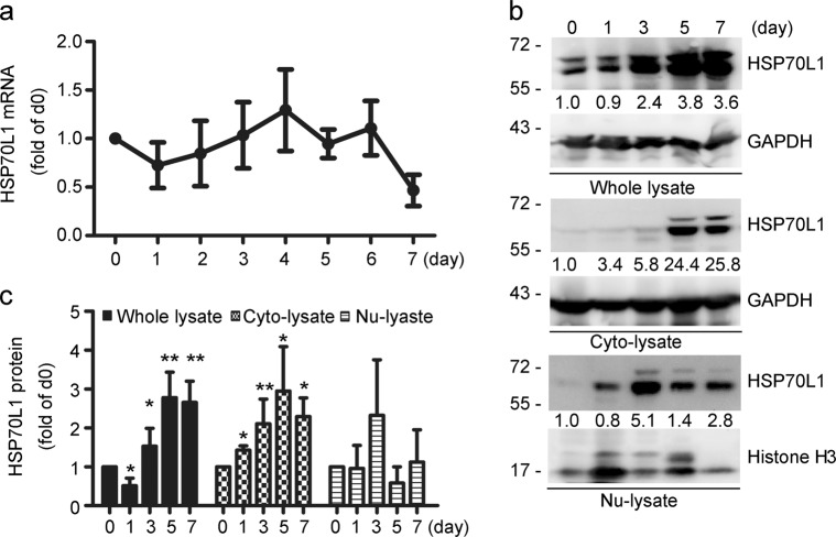
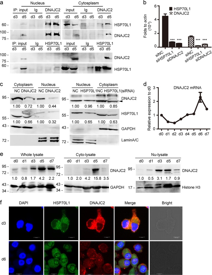
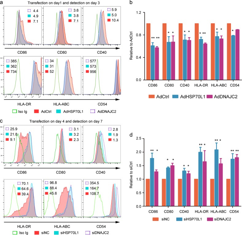
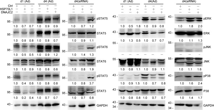
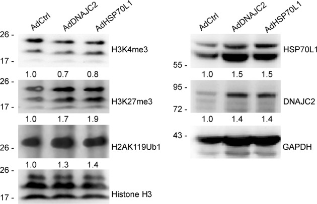
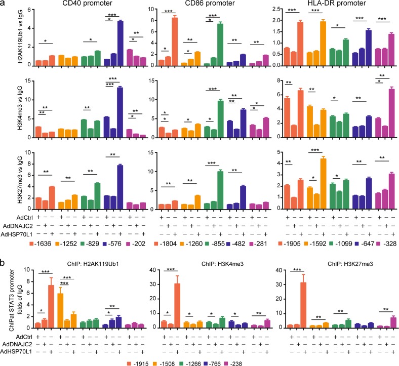
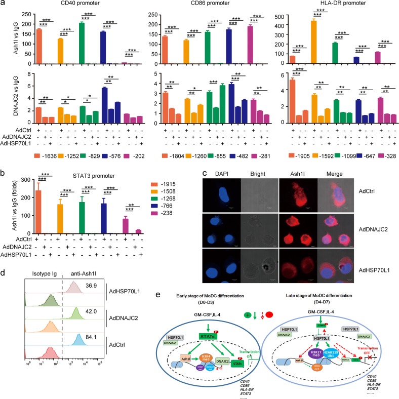
Epigenetic regulation has been attracting increasing attention due to its role in cell differentiation and behaviors. However, the epigenetic mechanisms that regulate human dendritic cell (DC) differentiation and development remain poorly understood. Our previous studies show that extracellular heat shock protein 70-like protein (HSP70L1) is a potent adjuvant of Th1 responses via stimulating DCs when released from cells; however, the role of intracellular HSP70L1 in DC differentiation and maturation remains unknown. Herein, we demonstrate that intracellular HSP70L1 inhibits human DC maturation by suppressing MHC and costimulatory molecule expression, in contrast to the adjuvant activity of extracellular HSP70L1. The stability of intracellular HSP70L1 is dependent on DNAJC2, a known epigenetic regulator. Mechanistically, intracellular HSP70L1 inhibits the recruitment of Ash1l to and maintains the repressive H3K27me3 and H2AK119Ub1 modifications on the promoter regions of costimulatory, MHC and STAT3 genes. Thus, intracellular HSP70L1 is an inhibitor of human DC maturation. Our results provide new insights into the epigenetic regulation of cell development by intracellular HSP70L1.

Keywords: DNAJC2; H2AK119Ub1; H3K27me3; H3K4me3; HSP70L1; Histone modification; Monocyte-derived dendritic cell.


Conflict of interest statement

The authors declare no competing interests.

Figures

Fig. 1
Fig. 1
HSP70L1 expression is dynamic during the differentiation of MoDCs. ac Real-time PCR (a) and WB (b, c) analyses of the mRNA and protein amounts of HSP70L1 in whole cell, nuclear, or cytoplasmic lysates from MoDCs, respectively, at indicated times during the differentiation of monocytes into DCs induced by GM-CSF and IL-4 for 7 days. Data are normalized to actin (a), GAPDH or Histone H3 (b, c), and day 0 group (monocytes) is set as 1. Data are shown as the mean±s.e.m. of three to five independent experiments (a, c), and one representative result is shown (b)
Fig. 2
Fig. 2
HSP70L1 and DNAJC2 interact with and stabilize each other. a Coimmunoprecipitation analysis of the interaction between HSP70L1 and DNAJC2 at indicated time during the differentiation of MoDCs. b, c Real-time PCR (b) and WB (c) analyses of the expression of HSP70L1 and DNAJC2 in MoDCs transfected with siRNAs of control (siNC), HSP70L1 (siHSP70L1) or DNAJC2 (siDNAJC2) on day 4 for 48 h (PCR) or 72 h (WB), respectively. Data are normalized to actin (b), GAPDH (c, cytoplasmic lysate), or LaminA/C (c, nuclear lysate), and siNC group is set as 1 (c). d, e Real-time PCR (d) and WB (e) analyses of the expression of DNAJC2 at indicated time during the differentiation of MoDCs. Data are normalized to GAPDH (d, e, whole cell and cytoplasmic lysate) or Histone H3 (e, nuclear lysate), and day 0 group is set as 1. f Confocal microscopy analysis of the expression of HSP70L1 and DNAJC2 in MoDCs on days 3 and 6, respectively (TCS-SP8, Leica, Wetzlar, Germany). Bars are indicated in figures. Data are shown as the mean ±s.e.m. of three experiments (b, d). One representative result of two (a, f) or three (c, e) independent experiments is shown. ***P < 0.001 compared to siNC group (t test)
Fig. 3
Fig. 3
HSP70L1 and DNAJC2 inhibit the maturation of MoDCs. ad The expression of costimulatory and MHC molecules was detected using FACS on MoDCs transfected with control Ad (AdCtrl), AdHSP70L1, or AdDNAJC2 on day 1 (a, b) or with siRNAs of control (siNC), HSP70L1 or DNAJC2 on day 4 (c, d). Numbers in histograms (a, c) that represent one of three independent experiments are relative fluorescence intensities to isotype IgG staining (a, c), and data in (b, d) are shown as the mean±s.e.m of three experiments. *p < 0.05, **p < 0.01 compared to AdCtrl or siNC group (t test)
Fig. 4
Fig. 4
HSP70L1 and DNAJC2 inhibit the activation of the STAT3, STAT5, ERK, and P38 pathways in MoDCs. The phosphorylation and constitutive expression of STAT3, STAT5, STAT6, ERK, P38, and JNK were detected after 36 h of transfection of MoDCs with AdCtrl, AdHSP70L1, or AdDNAJC2, respectively, on days 1 and 4, or 48 h of transfection of MoDCs with siNC, siHSP70L1, or siDNAJC2 on day 4. Gray intensities were normalized to GAPDH (constitutive expression) or corresponding non-phosphorylated transcription factors, and AdCtrl or siNC group was set as 1. Representative results of three independent experiments are shown
Fig. 5
Fig. 5
Overexpression of HSP70L1 and DNAJC2 enhances H3K27me3 and inhibits H3K4me3 in MoDCs. WB analysis of the H2AK119Ub1, H3K4me3, and H3K27me3 modifications in MoDCs after 48 h of transfection with AdCtrl, AdDNAJC2, or AdHSP70L1 on day 1. Gray intensities are normalized to Histone H3 or GAPDH, and AdCtrl group is set as 1. Representative results of three (right panel) or six (left panel) independent experiments are shown
Fig. 6
Fig. 6
Effects of the overexpression of HSP70L1 and DNAJC2 on the H2AK119Ub1, H3K4me3, and H3K27me3 modifications of the CD40, CD86, HLA-DR, and STAT3 promoters in MoDCs at early stage. ChIP analysis of the H2AK119Ub1, H3K4me3, and H3K27me3 modifications on the CD40, CD86, HLA-DR, and STAT3 promoter regions after 48 h of MoDCs transfected with AdCtrl, AdDNAJC2, or AdHSP70L1 on day 1. Data are normalized to input and then control IgG is set as 1. Data are shown as the mean ± s.d. of three determinants in one ChIP experiment. Representative results of three independent experiments are shown. *p < 0.05, **p < 0.01, ***p < 0.001 compared to AdCtrl group (t test)
Fig. 7
Fig. 7
Effects of the overexpression of HSP70L1 and DNAJC2 on the recruitment of Ash1l to the CD40, CD86, HLA-DR, and STAT3 promoters in MoDCs. a, b ChIP analysis of the recruitment of Ash1l (a, b) and DNAJC2 (a) to the CD40, CD86, HLA-DR, and STAT3 promoter regions after 48 h of MoDCs transfected with AdCtrl, AdDNAJC2, or AdHSP70L1 on day 1. Data are normalized to input, and control IgG is set as 1. Data are shown as the mean ± s.d. of three determinants in one ChIP. Representative results of three (a) or two (b) independent experiments are shown. *p < 0.05, **p < 0.01, ***p < 0.001 compared to AdCtrl group (t-test). (c, d) Confocal microscopy (c) and flow cytometry (d) analyses of the expression of Ash1l in MoDCs after 48 h of transfection with AdCtrl, AdDNAJC2, or AdHSP70L1 on day 1. Numbers in histograms that represent one of two independent experiments are relative fluorescence intensities to isotype IgG staining (d). e A mechanistic scheme of intracellular HSP70L1 inhibiting the maturation of MoDCs

References

    1. Hammer GE, Ma A. Molecular control of steady-state dendritic cell maturation and immune homeostasis. Annu. Rev. Immunol. 2013;31:743–791. - PMC - PubMed
    1. Worbs T, Hammerschmidt SI, Forster R. Dendritic cell migration in health and disease. Nat. Rev. Immunol. 2017;17:30–48. - PubMed
    1. Merad M, Sathe P, Helft J, Miller J, Mortha A. The dendritic cell lineage: ontogeny and function of dendritic cells and their subsets in the steady state and the inflamed setting. Annu. Rev. Immunol. 2013;31:563–604. - PMC - PubMed
    1. Mildner A, Jung S. Development and function of dendritic cell subsets. Immunity. 2014;40:642–656. - PubMed
    1. Pearce EJ, Everts B. Dendritic cell metabolism. Nat. Rev. Immunol. 2015;15:18–29. - PMC - PubMed

Publication types

MeSH terms