

Is CT or MRI the optimal imaging investigation for the diagnosis of large vestibular aqueduct syndrome and large endolymphatic sac anomaly?
- PMID: 30635710
- PMCID: PMC6411674
- DOI: 10.1007/s00405-019-05279-x
Is CT or MRI the optimal imaging investigation for the diagnosis of large vestibular aqueduct syndrome and large endolymphatic sac anomaly?
Abstract
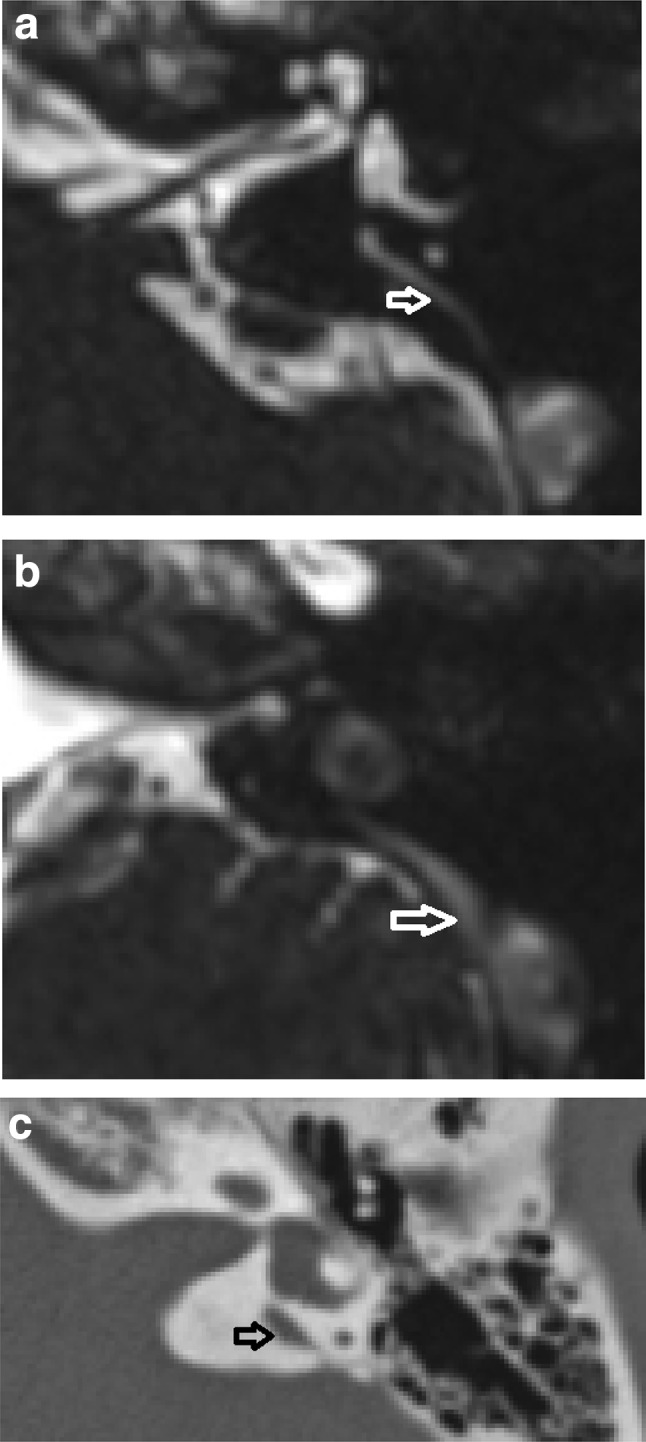
Background and purpose: We explored whether there was a difference between measurements obtained with CT and MRI for the diagnosis of large vestibular aqueduct syndrome or large endolymphatic sac anomaly, and whether this influenced diagnosis on the basis of previously published threshold values (Valvassori and Cincinnati). We also investigated whether isolated dilated extra-osseous endolymphatic sac occurred on MRI. Secondary objectives were to compare inter-observer reproducibility for the measurements, and to investigate any mismatch between the diagnoses using the different criteria.
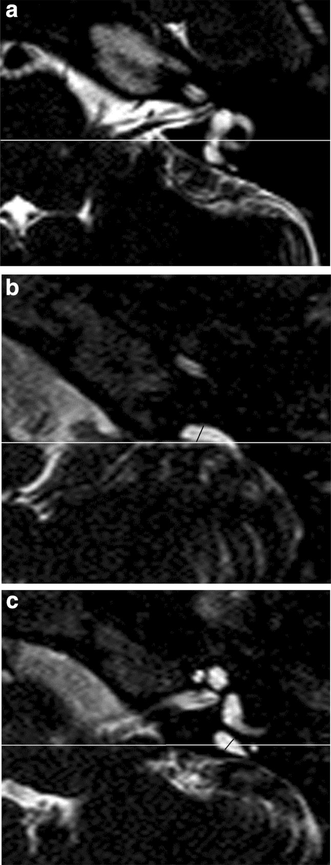
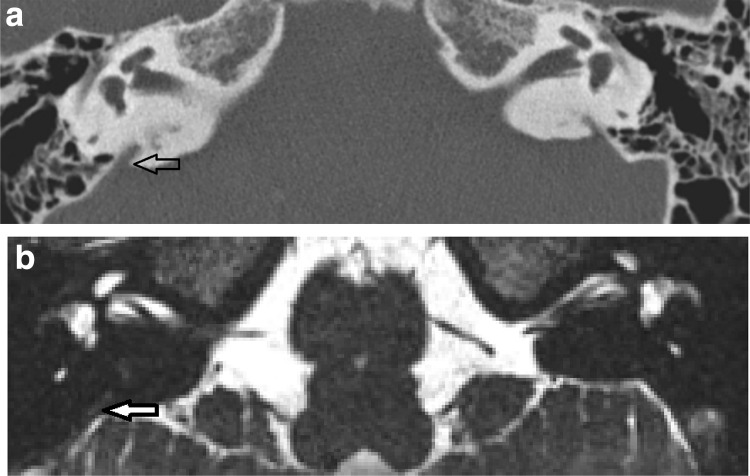
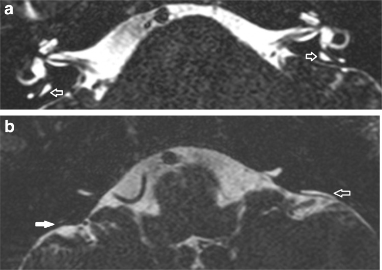
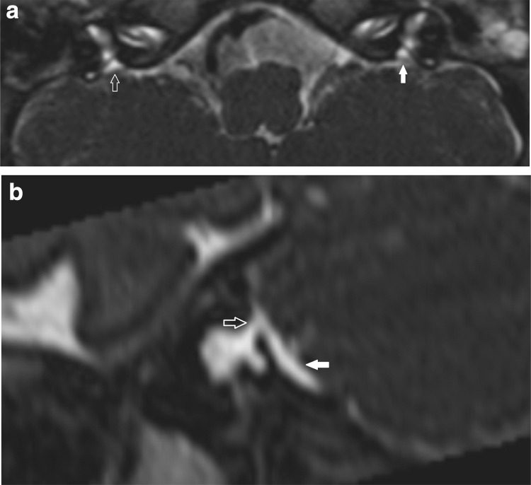
Materials/methods: Subjects diagnosed with large vestibular aqueduct syndrome or large endolymphatic sac anomalies were retrospectively analysed. For subjects with both CT and MRI available (n = 58), two independent observers measured the midpoint and operculum widths. For subjects with MRI (± CT) available (n = 84), extra-osseous sac widths were also measured. Results There was no significant difference between the width measurements obtained with CT versus MRI. CT alone diagnosed large vestibular aqueduct syndrome or large endolymphatic sac anomalies in 2/58 (Valvassori) and 4/58 (Cincinnati), whilst MRI alone diagnosed them in 2/58 (Valvassori). There was 93% CT/MRI diagnostic agreement using both criteria. Only 1/84 demonstrated isolated extra-osseous endolymphatic sac dilatation. The MRI-based LVAS/LESA diagnosis was less dependent on which criteria were used. Midpoint measurements are more reproducible between observers and between CT/MR imaging modalities.
Conclusion: Supplementing MRI with CT results in additional diagnoses using either criterion, however, there is no net increased diagnostic sensitivity for CT versus MRI when applying the Valvassori criteria. Isolated enlargement of the extra-osseous endolymphatic sac is rare.
Keywords: Computed tomography; Deafness; Inner ear; Large endolymphatic sac anomaly; Large vestibular aqueduct syndrome; Magnetic resonance imaging.
Conflict of interest statement
Conflict of interest
All authors, Connor SEJ, Dudau C, Pai I, and Gaganasiou M, declare that they have no conflict of interest.
Ethical standards
All procedures performed in studies involving human participants were in accordance with the ethical standards of the institutional and/or national research committee and with the 1964 Helsinki declaration and its later amendments or comparable ethical standards.
Informed consent
The study underwent local institutional review and was considered to represent service evaluation without a requirement for informed consent.
Figures
Similar articles
-
[Imaging characteristics of patients with large vestibular aqueduct syndrome and its relationship with the acoustically evoked short latency negative response].Zhonghua Er Bi Yan Hou Tou Jing Wai Ke Za Zhi. 2019 Aug 7;54(8):561-565. doi: 10.3760/cma.j.issn.1673-0860.2019.08.001. Zhonghua Er Bi Yan Hou Tou Jing Wai Ke Za Zhi. 2019. PMID: 31434367 Chinese.
-
Comparative Analysis of CT and MRI Diagnosis of Large Vestibular Aqueduct Syndrome (LVAS) in Children.J Coll Physicians Surg Pak. 2019 Aug;29(8):753-756. doi: 10.29271/jcpsp.2019.08.753. J Coll Physicians Surg Pak. 2019. PMID: 31358098
-
Evaluation of the radiological criteria to diagnose large vestibular aqueduct syndrome.Int J Pediatr Otorhinolaryngol. 2016 Feb;81:84-91. doi: 10.1016/j.ijporl.2015.12.012. Epub 2015 Dec 30. Int J Pediatr Otorhinolaryngol. 2016. PMID: 26810296
-
Large vestibular aqueduct syndrome with massive endolymphatic sacs.Otolaryngol Head Neck Surg. 1995 Nov;113(5):606-10. doi: 10.1177/019459989511300513. Otolaryngol Head Neck Surg. 1995. PMID: 7478652 Review. No abstract available.
-
Abnormalities, congenital anomalies, and unusual anatomic variations of the endolymphatic sac and vestibular aqueduct: clinical, surgical, and radiographic correlations.Am J Otol. 1980 Oct;2(2):118-49. Am J Otol. 1980. PMID: 7011045 Review. No abstract available.
Cited by
-
Does CISS MRI Reliably Depict the Endolymphatic Duct in Children with and without Vestibular Aqueduct Enlargement?AJNR Am J Neuroradiol. 2024 Apr 8;45(4):511-517. doi: 10.3174/ajnr.A8158. AJNR Am J Neuroradiol. 2024. PMID: 38423746 Free PMC article.
-
Rare Disorders of the Vestibular Labyrinth: of Zebras, Chameleons and Wolves in Sheep's Clothing.Laryngorhinootologie. 2021 Apr;100(S 01):S1-S40. doi: 10.1055/a-1349-7475. Epub 2021 Apr 30. Laryngorhinootologie. 2021. PMID: 34352900 Free PMC article. Review.
-
Can MRI biomarkers for hearing loss in enlarged vestibular aqueduct be measured reproducibly?Br J Radiol. 2023 Jul;96(1147):20220274. doi: 10.1259/bjr.20220274. Epub 2023 May 10. Br J Radiol. 2023. PMID: 37162001 Free PMC article.
-
A novel genotyping technique for discriminating LVAS-associated high-frequency variants in SLC26A4 gene.AMB Express. 2020 Sep 15;10(1):166. doi: 10.1186/s13568-020-01102-7. AMB Express. 2020. PMID: 32930899 Free PMC article.
-
Perilymphatic Fistula: A Review of Classification, Etiology, Diagnosis, and Treatment.Front Neurol. 2020 Sep 15;11:1046. doi: 10.3389/fneur.2020.01046. eCollection 2020. Front Neurol. 2020. PMID: 33041986 Free PMC article. Review.
References
-
- Emmett JR. The large vestibular aqueduct syndrome. Am J Otol. 1985;6:387–415. - PubMed
-
- Vijayasekaran S, Halstead MJ, Boston M, Meinzen-Derr J, Bardo DME, Greinwald J, Benton C. When is the vestibular aqueduct enlarged? A statistical analysis of the normative distribution of vestibular aqueduct size. AJNR Am J Neuroradiol. 2007;28:1133–1138. doi: 10.3174/ajnr.A0495. - DOI - PMC - PubMed
MeSH terms
LinkOut - more resources
Full Text Sources
Medical
