

Cerebral torque is human specific and unrelated to brain size
- PMID: 30635713
- PMCID: PMC6499874
- DOI: 10.1007/s00429-018-01818-0
Cerebral torque is human specific and unrelated to brain size
Abstract
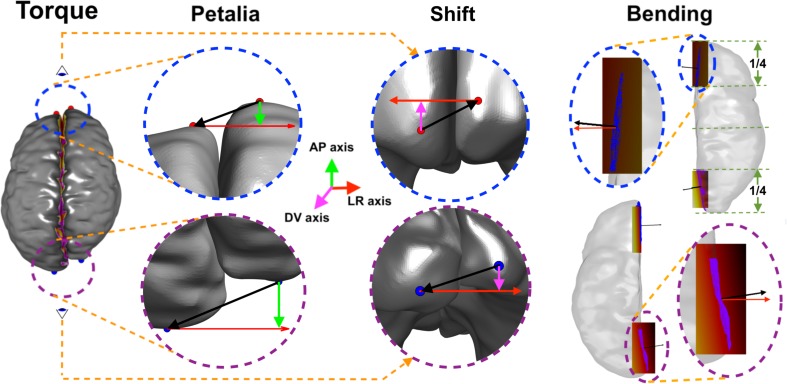
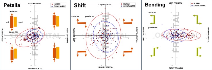
The term "cerebral torque" refers to opposing right-left asymmetries of frontal and parieto-occipital regions. These are assumed to derive from a lateralized gradient of embryological development of the human brain. To establish the timing of its evolution, we computed and compared the torque, in terms of three principal features, namely petalia, shift, and bending of the inter-hemispheric fissure as well as the inter-hemispheric asymmetry of brain length, height and width for in vivo Magnetic Resonance Imaging (MRI) scans of 91 human and 78 chimpanzee brains. We found that the cerebral torque is specific to the human brain and that its magnitude is independent of brain size and that it comprises features that are inter-related. These findings are consistent with the concept that a "punctuational" genetic change of relatively large effect introduced lateralization in the hominid lineage. The existence of the cerebral torque remains an unsolved mystery and the present study provides further support for this most prominent structural brain asymmetry being specific to the human brain. Establishing the genetic origins of the torque may, therefore, have relevance for a better understanding on human evolution, the organisation of the human brain, and, perhaps, also aspects of the neural basis of language.
Keywords: Asymmetry; Cerebral torque; Chimpanzee; Magnetic Resonance Imaging (MRI); Speciation.
Conflict of interest statement
Ethical approval
This manuscript is complied with Ethical Standards. All authors have been personally and actively involved in substantive work leading to the manuscript, and will hold themselves jointly and individually responsible for its content. Ethical approval was obtained from the local Research Ethics Committee where brain images were scanned.
Conflict of interest
We have no conflicts of interest to disclose and confirm that the manuscript is not under consideration for publication elsewhere.
Figures
References
-
- Annett M. Left, right, hand and brain: the right shift theory. London: Lawrence Erlbaum Associates; 1985.
-
- Balzeau A, Gilissen E. Endocranial shape asymmetries in pan paniscus, pan troglodytes and gorilla gorilla assessed via skull based landmark analysis. J Hum Evol. 2010;59:54–69. - PubMed
-
- Barrick TR, Mackay CE, Prima S, Maes F, Vandermeulen D, Crow TJ, Roberts N. Automatic analysis of cerebral asymmetry: an exploratory study of the relationship between brain torque and planum temporale asymmetry. NeuroImage. 2005;24(3):678–691. - PubMed
-
- Bear D, Schiff D, Saver J, Greenberg M, Freeman R. Quantitative analysis of cerebral asymmetries. Fronto-occipital correlation, sexual dimorphism and association with handedness. Arch Neurol. 1986;43(6):598–603. - PubMed
MeSH terms
LinkOut - more resources
Full Text Sources
