. 2019 Jan 11;114(2):7.
doi: 10.1007/s00395-018-0713-y.

Inhibition of cardiomyocyte Sprouty1 protects from cardiac ischemia-reperfusion injury

Affiliations

Inhibition of cardiomyocyte Sprouty1 protects from cardiac ischemia-reperfusion injury

Tarja Alakoski et al. Basic Res Cardiol. .

Abstract

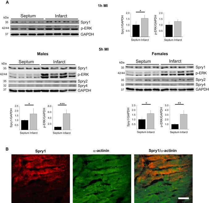
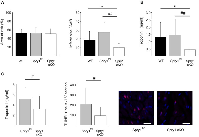
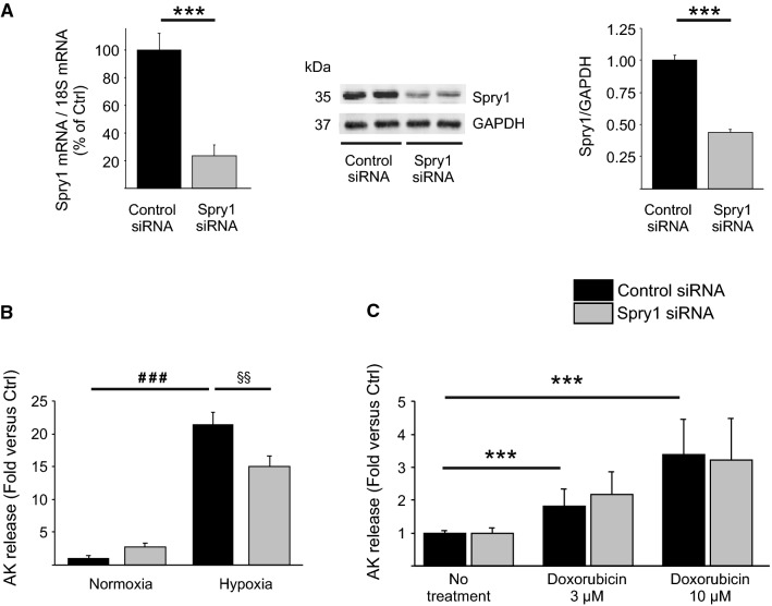
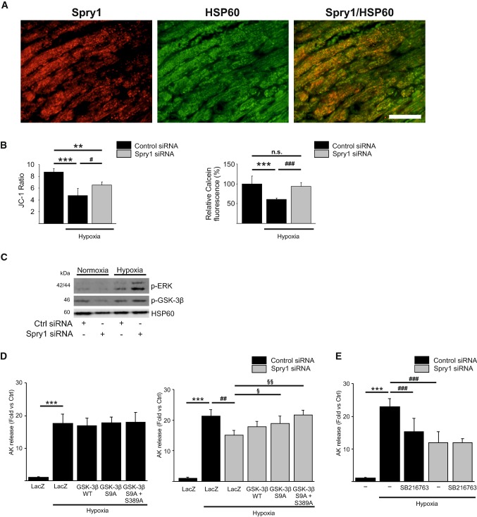
Sprouty1 (Spry1) is a negative modulator of receptor tyrosine kinase signaling, but its role in cardiomyocyte survival has not been elucidated. The aim of this study was to investigate the potential role of cardiomyocyte Spry1 in cardiac ischemia-reperfusion (I/R) injury. Infarct areas of mouse hearts showed an increase in Spry1 protein expression, which localized to cardiomyocytes. To investigate if cardiomyocyte Spry1 regulates I/R injury, 8-week-old inducible cardiomyocyte Spry1 knockout (Spry1 cKO) mice and control mice were subjected to cardiac I/R injury. Spry1 cKO mice showed reduction in release of cardiac troponin I and reduced infarct size after I/R injury compared to control mice. Similar to Spry1 knockdown in cardiomyocytes in vivo, RNAi-mediated Spry1 silencing in isolated cardiomyocytes improved cardiomyocyte survival following simulated ischemia injury. Mechanistically, Spry1 knockdown induced cardiomyocyte extracellular signal-regulated kinase (ERK) phosphorylation in healthy hearts and isolated cardiomyocytes, and enhanced ERK phosphorylation after I/R injury. Spry1-deficient cardiomyocytes showed better preserved mitochondrial membrane potential following ischemic injury and an increase in levels of phosphorylated ERK and phosphorylated glycogen synthase kinase-3β (GSK-3β) in mitochondria of hypoxic cardiomyocytes. Overexpression of constitutively active GSK-3β abrogated the protective effect of Spry1 knockdown. Moreover, pharmacological inhibition of GSK-3β protected wild-type cardiomyocytes from cell death, but did not further protect Spry1-silenced cardiomyocytes from hypoxia-induced injury. Cardiomyocyte Spry1 knockdown promotes ERK phosphorylation and offers protection from I/R injury. Our findings indicate that Spry1 is an important regulator of cardiomyocyte viability during ischemia-reperfusion injury.

Keywords: Extracellular signal-regulated kinase; Glycogen synthase kinase-3β; Ischemia–reperfusion injury; Myocardial infarction; Sprouty1.


Conflict of interest statement

The authors have no conflict of interest.

Figures

Fig. 1
Fig. 1
Analysis for Spry1 expression in ischemic myocardium. a Wild-type male mice were subjected to myocardial infarction (MI) for 1 and 5 h, and female mice for 5 h. Heart tissues from infarcted area and remote area (septum) were collected. Shown is immunoblot analysis of Spry1, phosphorylated extracellular signal-regulated kinase (p-ERK), Spry2, Spry4 and glyceraldehyde 3-phosphate dehydrogenase (GAPDH). Quantification of immunoblot analysis of Spry1 and p-ERK. GAPDH was used as a loading control. Data are shown as fold versus septum. N = 4–5; *P < 0.05, **P < 0.01, ***P < 0.001 versus septum. b Spry1fl/fl mice were subjected to ischemia–reperfusion (I/R) injury and heart tissue was collected after 6 h of reperfusion. Shown is the immunofluorescence staining of formalin fixed and paraffin-embedded tissue sections from infarct area for Spry1 and cardiomyocyte marker α-actinin. Scale bar 40 µm
Fig. 2
Fig. 2
Spry1 cardiomyocyte knockdown protects from ischemia–reperfusion injury in vivo. Wild-type (WT), Spry1fl/fl and Spry1 cardiomyocyte knockout (cKO) mice were subjected to 30 min of cardiac ischemia and 24 h of reperfusion, and subjected to analysis of infarct size, serum troponin I levels and analysis of cardiac function by echocardiography. a Analysis for area at risk (AAR) and infarct size relative to AAR derived from triphenyltetrazolium chloride (TTC) stainings. N = 9–13 per group. b Analysis for troponin I levels from serum samples. N = 9–13 per group. c Spry1fl/fl and Spry1 cKO mice were subjected to 30 min of ischemia and 6 h of reperfusion, and subjected to analysis for serum troponin I levels and apoptosis by terminal deoxynucleotidyl transferase dUTP nick end labeling (TUNEL). Shown are also representative images of TUNEL labeling of sections from hearts of Spry1fl/fl and Spry1 cKO mice. Scale bar 40 µm. N = 11–13 per group. *P < 0.05 versus WT mice and #P < 0.05, ##P < 0.01 versus Spry1fl/fl mice
Fig. 3
Fig. 3
Spry1 knockdown in isolated cardiomyocytes protects from hypoxia–reperfusion injury. Neonatal rat ventricular cardiomyocytes (CMs) were transfected with Spry1 siRNA (100 nM) or control siRNA (100 nM). a Three days later, RNA samples were collected from CMs and analyzed by qPCR. Shown is the relative expression of Spry1 mRNA in Spry1 RNAi treated and control RNAi treated CMs. Results were normalized to expression of 18S ribosomal RNA (18S). N = 3 for each group. Four days later, protein samples were collected from CMs and analyzed by western blotting. Shown is immunoblot analysis for Spry1 and glyceraldehyde 3-phosphate dehydrogenase (GAPDH) and quantification of immunoblot analysis of Spry1. GAPDH was used as a loading control. Data are shown as fold versus control RNAi. N = 3–6. b RNAi-treated CMs were subjected to hypoxia. Shown is the analysis for released adenylate kinase (AK) from damaged CMs after 4 h of hypoxia. N = 4 for each group. c RNAi-treated CMs were treated with 3 μM and 10 μM doxorubicin for 24 h. Shown is the analysis for AK release from damaged CMs. N = 3 for each group. ***P < 0.001 versus control RNAi; ###P < 0.001 versus normoxia control RNAi and §§P < 0.01 versus control RNAi in hypoxia
Fig. 4
Fig. 4
Spry1 regulates ERK and p38 in cardiomyocytes. Spry1fl/fl and Spry1 cardiomyocyte knockout (cKO) mice were subjected to sham operation or to 30 min of ischemia (I/R), and heart tissue was collected after 6 h of reperfusion. a Immunoblot analysis of phosphorylated extracellular signal-regulated kinase (p-ERK) and glyceraldehyde 3-phosphate dehydrogenase (GAPDH) in sham and I/R operated Spry1fl/fl and Spry1 cKO hearts. b Neonatal rat ventricular cardiomyocytes were transfected with Spry1 siRNA (100 nM) or control siRNA (100 nM). Four days later, protein samples were collected. Shown is the immunoblot analysis for Spry1, p-ERK and glyceraldehyde 3-phosphate dehydrogenase (GAPDH). c Immunofluorescence analysis for p-ERK and F-actin marker Phalloidin from control hearts and infarct areas from hearts subjected to 6 h of I/R injury. Scale bar 40 µm. d Immunoblot analysis of phosphorylated p38 (p-p38) and GAPDH in sham and I/R operated Spry1fl/fl and Spry1 cKO hearts. e Immunoblot analysis for Spry1, p-p38 and GAPDH from protein samples of neonatal rat ventricular cardiomyocytes transfected with Spry1 siRNA (100 nM) or control siRNA (100 nM)
Fig. 5
Fig. 5
Glycogen synthase kinase-3β mediates the protective effect of Spry1 knockdown. a Spry1fl/fl mice were subjected to 30 min of ischemia and heart tissue was collected after 6 h of reperfusion. Shown is the immunofluorescence staining of frozen sections from infarct area for Spry1 and mitochondrial marker heat shock protein 60 (HSP60). Scale bar 40 µm. b Neonatal rat ventricular cardiomyocytes (CMs) were transfected with Spry1 siRNA (100 nM) or control siRNA (100 nM), and 4 days later subjected to hypoxia for 4 h. Shown is the analysis for mitochondrial membrane potential expressed as ratio of JC-1 aggregate versus JC-1 monomer and detection of mitochondrial permeability transition pore opening using calcein cobalt assay expressed as relative Calcein-AM fluorescence (%). N = 4–8; **P < 0.01, ***P < 0.001 versus normoxia control RNAi, #P < 0.05, ###P < 0.001 versus hypoxia control RNAi. c RNAi treated CMs were subjected to hypoxia for 4 h and mitochondrial protein samples were collected and analyzed by western blotting for phosphorylated extracellular signal-regulated kinase (p-ERK), phosphorylated glycogen synthase kinase-3β (p-GSK-3β) and HSP60. d RNAi-treated CMs were transduced with adenoviruses expressing LacZ, wild-type (WT) GSK-3β, Ser9Ala GSK-3β or Ser9Ala/Ser389Ala double mutant GSK-3β, and subjected to hypoxia for 4 h. Shown is the analysis for released adenylate kinase (AK) from damaged CMs after hypoxia. N = 4 for each group; ***P < 0.001 versus normoxia control RNAi, ##P < 0.01 versus hypoxia control RNAi and §P < 0.05, §§P < 0.01 versus hypoxia Spry1 RNAi. e RNAi-treated CMs were treated with GSK-3β inhibitor SB216763 (3 μM) for 20 min and subjected to hypoxia for 4 h. Shown is the analysis for released AK from damaged CMs after hypoxia. N = 4 each group; ***P < 0.001 versus normoxia control RNAi, ###P < 0.001 versus hypoxia control RNAi

References

    1. Anderson K, Nordquist KA, Gao X, Hicks KC, Zhai B, Gygi SP, Patel TB. Regulation of cellular levels of Sprouty2 protein by prolyl hydroxylase domain and von Hippel–Lindau proteins. J Biol Chem. 2011;286:42027–42036. doi: 10.1074/jbc.M111.303222. - DOI - PMC - PubMed
    1. Argaud L, Gateau-Roesch O, Raisky O, Loufouat J, Robert D, Ovize M. Postconditioning inhibits mitochondrial permeability transition. Circulation. 2005;111:194–197. doi: 10.1161/01.CIR.0000151290.04952.3B. - DOI - PubMed
    1. Basson MA, Akbulut S, Watson-Johnson J, Simon R, Carroll TJ, Shakya R, Gross I, Martin GR, Lufkin T, McMahon AP, Wilson PD, Costantini FD, Mason IJ, Licht JD. Sprouty1 is a critical regulator of GDNF/RET-mediated kidney induction. Dev Cell. 2005;8:229–239. doi: 10.1016/j.devcel.2004.12.004. - DOI - PubMed
    1. Beurel E, Grieco SF, Jope RS. Glycogen synthase kinase-3 (GSK3): regulation, actions, and diseases. Pharmacol Ther. 2015;148:114–131. doi: 10.1016/j.pharmthera.2014.11.016. - DOI - PMC - PubMed
    1. Botker HE, Hausenloy D, Andreadou I, Antonucci S, Boengler K, Davidson SM, Deshwal S, Devaux Y, Di Lisa F, Di Sante M, Efentakis P, Femmino S, Garcia-Dorado D, Giricz Z, Ibanez B, Iliodromitis E, Kaludercic N, Kleinbongard P, Neuhauser M, Ovize M, Pagliaro P, Rahbek-Schmidt M, Ruiz-Meana M, Schluter KD, Schulz R, Skyschally A, Wilder C, Yellon DM, Ferdinandy P, Heusch G. Practical guidelines for rigor and reproducibility in preclinical and clinical studies on cardioprotection. Basic Res Cardiol. 2018;113:39. doi: 10.1007/s00395-018-0696-8. - DOI - PMC - PubMed

Publication types