. 2019 Oct;191(2):370-381.
doi: 10.1007/s12011-019-1631-z. Epub 2019 Jan 11.

Zinc Deficiency and Arsenic Exposure Can Act Both Independently or Cooperatively to Affect Zinc Status, Oxidative Stress, and Inflammatory Response

Affiliations

Zinc Deficiency and Arsenic Exposure Can Act Both Independently or Cooperatively to Affect Zinc Status, Oxidative Stress, and Inflammatory Response

Carmen P Wong et al. Biol Trace Elem Res. 2019 Oct.

Abstract

The negative health impact of zinc deficiency overlaps significantly with arsenic exposure, and is associated with increased risk for chronic diseases. Arsenic contamination in the groundwater often co-exists in regions of the world that are prone to zinc deficiency. Notably, low zinc status shares many hallmarks of arsenic exposure, including increased oxidative stress and inflammation. Despite their common targets and frequent co-distribution in the population, little is known regarding the interaction between zinc deficiency and arsenic exposure. In this study, we tested the effect of arsenic exposure at environmentally relevant doses in combination with a physiologically relevant level of zinc deficiency (marginal zinc deficiency) on zinc status, oxidative damage, and inflammation. In cell culture, zinc-deficient THP-1 monocytes co-exposed with arsenic resulted in further reduction in intracellular zinc, as well as further increase in oxidative stress and inflammatory markers. In an animal study, zinc-deficient mice had further decrease in zinc status when co-exposed to arsenic. Zinc deficiency, but not arsenic exposure, resulted in an increase in baseline transcript abundance of inflammatory markers in the liver. Upon lipopolysaccharide challenge to elicit an acute inflammatory response, arsenic exposure, but not zinc deficiency, resulted in an increase in proinflammatory response. In summary, zinc deficiency and arsenic exposure can function independently or cooperatively to affect zinc status, oxidant stress, and proinflammatory response. The results highlight the need to consider both nutritional status and arsenic exposures together when considering their impact on health outcomes in susceptible populations.

Keywords: Arsenic; Inflammation; Oxidative stress; Zinc.


Conflict of interest statement

Conflict of Interest

The authors declare that they have no conflict of interest.

Figures

Fig. 1
Fig. 1. Zinc deficiency enhanced arsenic-induced ROS production.
THP-1 cells were grown in zinc adequate (ZA) or zinc deficient (ZD) media for 4 weeks, followed by 24h arsenic exposure. Cells were labeled with H2DCFDA, a ROS indicator. Conversion of the nonfluorescent H2DCFDA to the highly fluorescent DCF in the presence of ROS were determined at 4h post-labeling. Data represent percent change compared to ZA control (0 μM As), n = 6–10 per group. Boxes represent interquartile range, the line inside each box represents the median, and whiskers represent maximum and minimum values, with individual data points shown as black filled circles. Two-way ANOVA to test for main effects of zinc status and arsenic treatment, followed by Bonferroni post-test for pairwise comparison: *** P<0.001, **** P<0.0001 compared to respective ZA group at different arsenic doses
Fig. 2
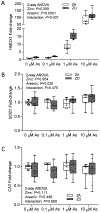
Fig. 2. Zinc deficiency increased arsenic-induced HMOX1 transcript abundance.
THP-1 cells were grown in zinc adequate (ZA) or zinc deficient (ZD) media for 4 weeks, followed by 4h arsenic exposure. Changes in transcript abundance of genes associated with oxidative stress were determined, including (a) HMOX1, (b) SOD1, and (c) CAT. Data represent normalized fold-change compared to ZA control of respective gene of interest (0 μM As), n = 8–12 per group. Boxes represent interquartile range, the line inside each box represents the median, and whiskers represent maximum and minimum values, with individual data points shown as black filled circles. Two-way ANOVA to test for main effects of zinc status and arsenic treatment, followed by Bonferroni post-test for pairwise comparison: **** P<0.0001 compared to respective ZA group at different arsenic doses
Fig. 3
Fig. 3. Arsenic exposure reduced intracellular zinc in THP-1 cells.
THP-1 cells were grown in zinc adequate (ZA) or zinc deficient (ZD) media for 4 weeks. Following 24h arsenic exposure, cells were (a) left untreated, or (b) treated with 10 ng/ml LPS for 24h prior to FluoZin3 labeling. Intracellular zinc levels were determined by flow cytometry. Data represent mean fluorescence intensity (MFI), n = 4 per group. Boxes represent interquartile range, the line inside each box represents the median, and whiskers represent maximum and minimum values, with individual data points shown as black filled circles. Two-way ANOVA to test for main effects of zinc status and arsenic treatment, followed by Bonferroni post-test for pairwise comparison: ** P<0.01, *** P<0.001 compared to respective 0 μM group in ZA or ZD treatment. Representative of three independent experiments
Fig. 4
Fig. 4. Arsenic and zinc deficiency enhanced proinflammatory response in THP-1 cells.
THP-1 cells were grown in zinc adequate (ZA) or zinc deficient (ZD) media for 4 weeks. In the top panel, (a) ICAM1, (b) IL6, and (c) CXCL8 transcript abundance were determined in ZA and ZD THP-1 cells after 24h arsenic treatment. In the bottom panel, ZA and ZD THP-1 cells were treated with arsenic for 24h, followed by 3h LPS (10 ng/ml) stimulation. LPS-induced changes in transcript abundance of (d) ICAM1, (e) IL6, and (f) CXCL8 were determined. Data represent normalized fold-change compared to ZA control of respective gene (0 μM As, no LPS), n = 7–12 per group. Boxes represent interquartile range, the line inside each box represents the median, and whiskers represent maximum and minimum values, with individual data points shown as black filled circles. Two-way ANOVA to test for main effects of zinc status and arsenic treatment, followed by Bonferroni post-test for pairwise comparison: ** P<0.01, *** P<0.001, **** P<0.0001 compared to respective ZA group with or without arsenic exposure
Fig. 5
Fig. 5. Zinc repletion did not restore zinc status or fully normalize zinc deficiency-enhanced oxidative stress and proinflammatory response.
THP-1 cells were grown in zinc adequate (ZA) or zinc deficient (ZD) media for 4 weeks. A subset of ZD THP-1 cells were then cultured for 3 days in ZA media to replete zinc prior to arsenic exposure. Intracellular zinc levels and transcript abundance in THP-1 cells were determined after 24h arsenic treatment. (a) Intracellular zinc levels were determined by FluoZin3 labeling. Data represent mean fluorescence intensity (MFI), n = 4 per group, representative of two independent experiments. Transcript abundance of (b) HMOX1, (c) ICAM1, and (d) IL6 were determined. Data in panel B-D represent normalized fold-change compared to ZA control of respective genes within each panel (0 μM As), n = 7–8 per group. Boxes represent interquartile range, the line inside each box represents the median, and whiskers represent maximum and minimum values, with individual data points shown as black filled circles. Two-way ANOVA to test for main effects of zinc status and arsenic treatment, followed by Bonferroni post-test for pairwise comparison: * P<0.05, ** P<0.01, *** P<0.001, **** P<0.0001 compared to respective ZA group with or without arsenic exposure
Fig. 6
Fig. 6. Arsenic reduced plasma zinc levels in mice.
C57Bl/6 mice were fed a zinc adequate diet (ZA, 30 mg/kg) or a marginal zinc deficient diet (MZD, 6mg/kg zinc), in combination with arsenic provided in the drinking water at 0, 50, or 500ppb. Plasma zinc levels were determined by ICP-OES after 6 wks dietary treatment, n = 7 per group. Boxes represent interquartile range, the line inside each box represents the median, and whiskers represent maximum and minimum values, with individual data points shown as black filled circles. Two-way ANOVA to test for main effects of zinc status and arsenic treatment, followed by Bonferroni post-test for pairwise comparison
Fig. 7
Fig. 7. Differential effects of zinc deficiency and arsenic exposure in increasing oxidative stress and proinflammatory response in mice.
C57Bl/6 mice were fed a zinc adequate diet (ZA, 30 mg/kg zinc) or a marginal zinc deficient diet (MZD, 6 mg/kg zinc), in combination with arsenic provided in the drinking water at 0, 50, or 500 ppb. In the first set of mice, changes in transcript abundance of genes associated with oxidative stress and inflammatory response in the liver were assessed after 6 wks dietary treatment (a-d). In a second set of mice, an acute proinflammatory response was induced by injecting mice i.p. with LPS (1 mg/g body weight) at the end of 6 wks dietary treatment (e-h). Liver samples were collected 3h post-LPS injections. Changes in the transcript abundance of Hmox1 (a and e), Il6 (b and d), Ccl2 (C and F), and Icam1 (d and g) were determined. Data represent normalized fold-change compared to ZA control of respective gene (ZA, 0 μM As, no LPS), n = 7 per group. Boxes represent interquartile range, the line inside each box represents the median, and whiskers represent maximum and minimum values, with individual data points shown as black filled circles. Two-way ANOVA to test for main effects of zinc status and arsenic treatment, followed by Bonferroni post-test for pairwise comparison: * P<0.05, ** P<0.01 compared to respective ZA group within each arsenic exposure

References

    1. Marreiro DD, Cruz KJ, Morais JB, Beserra JB, Severo JS, de Oliveira AR (2017) Zinc and Oxidative Stress: Current Mechanisms. Antioxidants (Basel) 6 (2). doi:10.3390/antiox6020024 - DOI - PMC - PubMed
    1. Prasad AS (2014) Zinc is an Antioxidant and Anti-Inflammatory Agent: Its Role in Human Health. Front Nutr 1:14. doi:10.3389/fnut.2014.00014 - DOI - PMC - PubMed
    1. Khansari N, Shakiba Y, Mahmoudi M (2009) Chronic inflammation and oxidative stress as a major cause of age-related diseases and cancer. Recent Pat Inflamm Allergy Drug Discov 3 (1):73–80 - PubMed
    1. Prasad AS (2009) Zinc: role in immunity, oxidative stress and chronic inflammation. Curr Opin Clin Nutr Metab Care 12 (6):646–652. doi:10.1097/MCO.0b013e3283312956 - DOI - PubMed
    1. Reuter S, Gupta SC, Chaturvedi MM, Aggarwal BB (2010) Oxidative stress, inflammation, and cancer: how are they linked? Free Radic Biol Med 49 (11):1603–1616. doi:10.1016/j.freeradbiomed.2010.09.006 - DOI - PMC - PubMed