. 2019 Jan 22;8(2):e010838.
doi: 10.1161/JAHA.118.010838.

Pretreatment With PCSK9 Inhibitor Protects the Brain Against Cardiac Ischemia/Reperfusion Injury Through a Reduction of Neuronal Inflammation and Amyloid Beta Aggregation

Affiliations

Pretreatment With PCSK9 Inhibitor Protects the Brain Against Cardiac Ischemia/Reperfusion Injury Through a Reduction of Neuronal Inflammation and Amyloid Beta Aggregation

Nattayaporn Apaijai et al. J Am Heart Assoc. .

Abstract

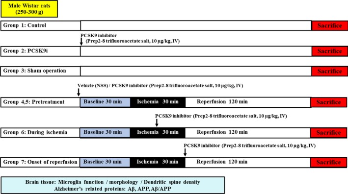
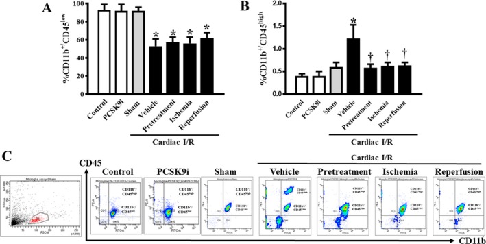
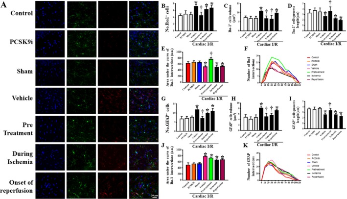
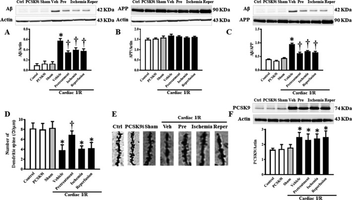
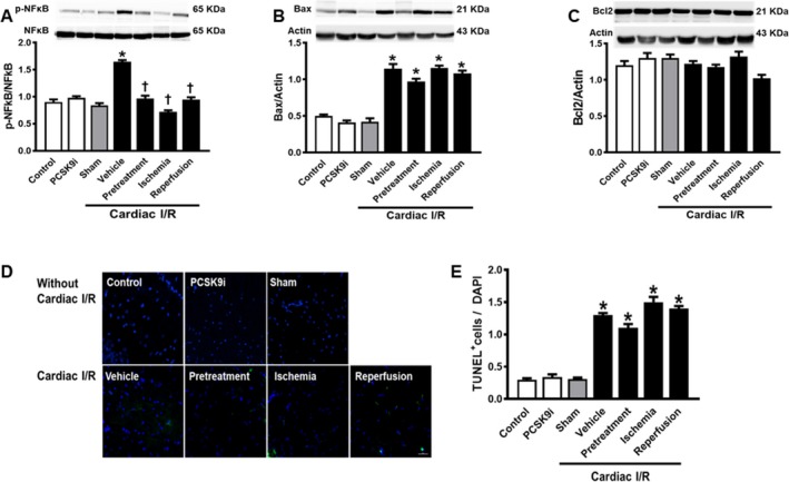
Background Cardiac ischemic/reperfusion (I/R) injury leads to brain damage. A new antihyperlipidemic drug is aimed at inhibiting PCSK 9 (proprotein convertase subtilisin/kexin type 9), a molecule first identified in a neuronal apoptosis paradigm. Thus, the PCSK 9 inhibitor ( PCSK 9i) may play a role in neuronal recovery following cardiac I/R insults. We hypothesize that PCSK 9i attenuates brain damage caused by cardiac I/R via diminishing microglial/astrocytic hyperactivation, β-amyloid aggregation, and loss of dendritic spine. Methods and Results Adult male rats were divided into 7 groups: (1) control (n=4); (2) PCSK 9i without cardiac I/R (n=4); (3) sham (n=4); and cardiac I/R (n=40). Cardiac I/R rats were divided into 4 subgroups (n=10/subgroup): (1) vehicle; (2) PCSK 9i (10 μg/kg, IV) before ischemia; (3) PCSK 9i during ischemia; and (4) PCSK 9i at the onset of reperfusion. At the end of cardiac I/R protocol, brains were removed to determine microglial and astrocytic activities, β-amyloid aggravation, and dendritic spine density. The cardiac I/R led to the activation of the brain's innate immunity resulting in increasing Iba1+ microglia, GFAP + astrocytes, and CD 11b+/ CD 45+high cell numbers. However, CD 11b+/ CD 45+low cell numbers were decreased following cardiac I/R. In addition, cardiac I/R led to reduced dendritic spine density, and increased β-amyloid aggregation. Only the administration of PCSK 9i before ischemia effectively attenuated these deleterious effects on the brain following cardiac I/R. PCSK 9i administration under the physiologic condition did not affect the aforementioned parameters. Conclusions Cardiac I/R injury activated microglial activity in the brain, leading to brain damage. Only the pretreatment with PCSK 9i prevented dendritic spine loss via reduction of microglial activation and Aβ aggregation.

Keywords: amyloid; brain; ischemia/reperfusion injury/neuroprotection; proprotein convertase subtilisin/kexin type 9.


Figures

Figure 1
Figure 1
Experimental protocol of the study. APP indicates amyloid beta precursor protein; Aβ, amyloid beta; PCSK9, proprotein convertase subtilisin/kexin type 9.
Figure 2
Figure 2
Effects of PCSK9 inhibition on microglial count after cardiac I/R injury. A, Percentage of CD11b+/CD45+ microglia of the brains. B, Percentage of CD11b+/CD45+high microglia of the brains. C, Representative flow cytometry images of microglial count. I/R, ischemia/reperfusion; CD, cluster of differentiation; PCSK9i, proprotein convertase subtilisin/kexin type 9 inhibitor; Control, rat group with no surgical intervention; PCSK9i, rats group with no intervention and treated with PCSK9i for 180 minutes; Sham, rat group with open chest surgery; Vehicle, rats treated with vehicle at 15 minutes before cardiac I/R; Pretreatment, rats treated with PCSK9i at 15 minutes before cardiac I/R; Ischemia, rats treated with PCSK9i at 15 minutes during ischemic period; Reperfusion, rats treated with PCSK9i at the onset of reperfusion period (n=4–10 per group). *P<0.05 vs control, PCSK9i, and sham, P<0.05 vs vehicle.
Figure 3
Figure 3
Effects of PCSK9 inhibition on microglial and astrocytic morphology after cardiac I/R injury. A, Representative confocal microscopy images of microglia (Iba1), astrocytes (GFAP), neuronal nuclei (DAPI), and all merged. Statistical analysis for the microglia fraction including: B, number of Iba1‐positive cells (units); C, dendrite volume (μm3); D, Iba1 filament dendrite length (μm); E, dendritic complexity as area under the curve of the Sholl analysis; F, dendritic complexity as slope of Sholl analysis; G, number of GFAP–positive cells (units); H, dendrite volume (μm3); I, GFAP filament dendrite length (μm); J, dendritic complexity as area under the curve of the Sholl analysis; K, dendritic complexity as slope of Sholl analysis. I/R, ischemia/reperfusion; Iba1, ionized calcium‐binding adapter molecule 1; GFAP, glial fibrillary acidic protein; PCSK9i, proprotein convertase subtilisin/kexin type 9 inhibitor; Control, rat group with no surgical intervention; PCSK9i, rats group with no intervention and treated with PCSK9i for 180 minutes; Sham, rat group with no surgical intervention/treatment; Vehicle, rats treated with vehicle at 15 minutes before cardiac I/R; Pretreatment, rats treated with PCSK9i at 15 minutes before cardiac I/R; Ischemia, rats treated with PCSK9i at 15 minutes during ischemic period; Reperfusion, rats treated with PCSK9i at the onset of reperfusion period (n=4–10 per group). *P<0.05 vs sham, P<0.05 vs vehicle.
Figure 4
Figure 4
Effects of PCSK9 inhibition on Aβ and APP protein expression levels and dendritic spine density after cardiac I/R injury. A, Protein levels of Aβ established by Western blotting, normalized to actin. B, Protein levels of APP by Western blotting, normalized to actin. C, Protein levels of Aβ by Western blotting, normalized to APP. D, Dendritic spine density per 20 μm apical tertiary dendrite; E, Representative images of dendritic spines. F, PCSK9 levels by Western blotting, normalized to actin. Aβ, amyloid beta; I/R, ischemia/reperfusion; Veh, vehicle; Pre, pretreatment; Reper, reperfusion; PCSK9, Proprotein convertase subtilisin/kexin type 9; PCSK9i, proprotein convertase subtilisin/kexin type 9 inhibitor; Control, rat group with no surgical intervention; PCSK9i, rats group with no intervention and treated with PCSK9i for 180 minutes; Sham, rat group with no surgical intervention/treatment; Vehicle, rats treated with vehicle at 15 minutes before cardiac I/R; Pretreatment, rats treated with PCSK9i at 15 minutes before cardiac I/R; Ischemia, rats treated with PCSK9i at 15 minutes during ischemic period; Reperfusion, rats treated with PCSK9i at the onset of reperfusion period (n=4–10 per group). *P<0.05 vs sham, P<0.05 vs vehicle.
Figure 5
Figure 5
Effects of PCSK9 inhibition on brain inflammation and apoptosis. A, p‐NFκB/NFκB established by Western blotting. B, Protein levels of Bax by Western blotting, normalized to actin. C, Protein levels of Bax by Western blotting, normalized to actin. D, Representative picture of TUNEL. E, TUNEL + cells/DAPI. I/R, ischemia/reperfusion; Veh, vehicle; Pre, pretreatment; Reper, reperfusion; TUNEL, terminal deoxynucleotidyl transferase dUTP nick end labeling; PCSK9i, Proprotein convertase subtilisin/kexin type 9 inhibitor; Control, rat group with no surgical intervention; PCSK9i, rats group with no intervention and treated with PCSK9i for 180 minutes; Sham, rat group with no surgical intervention/treatment; Vehicle, rats treated with vehicle at 15 minutes before cardiac I/R; Pretreatment, rats treated with PCSK9i at 15 minutes before cardiac I/R; Ischemia, rats treated with PCSK9i at 15 minutes during ischemic period; Reperfusion, rats treated with PCSK9i at the onset of reperfusion period (*P<0.05 vs. sham, P<0.05 vs. vehicle. n=4–10 per group).
Figure 6
Figure 6
A summary diagram showing the possible effects of PCSK9 inhibition on the brain following cardiac I/R injury. During cardiac I/R injury, the brain PCSK9 levels had increased, along with neuronal inflammation, and Aβ aggregation, leading to a reduction in dendritic spine density. Treatment with PCSK9i increased dendritic spine density by reducing neuronal inflammation and Aβ aggregation, but it did not affect the brain PCSK9 levels. Thus, we proposed that the beneficial effects of PCSK9i on the brain were possibly because of a reduction of systemic inflammation during cardiac I/R injury. Aβ indicates amyloid beta; I/R, ischemia/reperfusion; PCSK9i, proprotein convertase subtilisin/kexin type 9 inhibitor.

References

    1. Palmerini T, Tomasi L, Barozzi C, Della Riva D, Mariani A, Taglieri N, Leone O, Ceccarelli C, De Servi S, Branzi A, Genereux P, Stone GW, Ahamed J. Detection of tissue factor antigen and coagulation activity in coronary artery thrombi isolated from patients with ST‐segment elevation acute myocardial infarction. PLoS One. 2013;8:e81501. - PMC - PubMed
    1. Ramirez‐Sanchez J, Pires ENS, Meneghetti A, Hansel G, Nunez‐Figueredo Y, Pardo‐Andreu GL, Ochoa‐Rodríguez E, Verdecia‐Reyes Y, Delgado‐Hernández R, Salbego C, Souza DO. JM‐20 treatment after MCAO reduced astrocyte reactivity and neuronal death on peri‐infarct regions of the rat brain. Mol Neurobiol. 2018. Available at: https://link.springer.com/article/10.1007%2Fs12035-018-1087-8. Accessed January 3, 2019. - PubMed
    1. Ibanez B, James S, Agewall S, Antunes MJ, Bucciarelli‐Ducci C, Bueno H, Caforio ALP, Crea F, Goudevenos JA, Halvorsen S, Hindricks G, Kastrati A, Lenzen MJ, Prescott E, Roffi M, Valgimigli M, Varenhorst C, Vranckx P, Widimský P. 2017 ESC guidelines for the management of acute myocardial infarction in patients presenting with ST‐segment elevation. Rev Esp Cardiol (Engl Ed). 2017;70:1082. - PubMed
    1. Kumfu S, Charununtakorn ST, Jaiwongkam T, Chattipakorn N, Chattipakorn SC. Humanin prevents brain mitochondrial dysfunction in a cardiac ischaemia‐reperfusion injury model. Exp Physiol. 2016;101:697–707. - PubMed
    1. Nour M, Scalzo F, Liebeskind DS. Ischemia‐reperfusion injury in stroke. Interv Neurol. 2013;1:185–199. - PMC - PubMed

Publication types

LinkOut - more resources