High-grade serous ovarian carcinoma with mucinous differentiation: report of a rare and unique case suggesting transition from the "SET" feature of high-grade serous carcinoma to the "STEM" feature
- PMID: 30636633
- PMCID: PMC6330567
- DOI: 10.1186/s13000-019-0781-9
High-grade serous ovarian carcinoma with mucinous differentiation: report of a rare and unique case suggesting transition from the "SET" feature of high-grade serous carcinoma to the "STEM" feature
Abstract
Background: High-grade serous carcinoma, a representative high-grade ovarian carcinoma, is believed to be closely associated with a TP53 mutation. Recently, this category of ovarian carcinoma has gained increasing attention owing to the recognition of morphological varieties of TP53-mutated high-grade ovarian carcinoma. Herein, we report the case of a patient with high-grade serous carcinoma with mucinous differentiation.
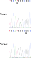
Case presentation: A 59-year-old postmenopausal woman was referred to the gynecologist because of abnormal vaginal bleeding. The radiological assessment revealed an intrapelvic multicystic mass, which was interpreted as an early right ovarian cancer and then removed by radical surgery. Histologically, the cancer cells were found in the bilateral ovaries and para-aortic lymph nodes. The cancer cells showed high-grade nuclear atypia and various morphologies, including the solid, pseudo-endometrioid, transitional cell-like (SET) pattern, and mucin-producing patterns. Benign and/or borderline mucin-producing epithelium, serous tubal intraepithelial carcinoma, and endometriosis-related lesions were not observed. In immunohistochemistry analyses, the cancer cells were diffuse positive for p53; block positive for p16; partial positive for WT1, ER, PgR, CDX2 and PAX8; and negative for p40, p63, GATA3, Napsin A, and vimentin. The Ki-67 labeling index of the cancer cells was 60-80%. Direct sequencing revealed that the cancer cells contained a missense mutation (c.730G>A) in the TP53 gene.
Conclusion: Mucinous differentiation in high-grade serous carcinoma is a rare and unique ovarian tumor phenotype and it mimics the phenotypes of mucinous or seromucinous carcinoma. To avoid the misdiagnosis, extensive histological and immunohistochemical analyses should be performed when pathologists encounter high-grade mucin-producing ovarian carcinoma. The present case shows that the unusual histological characteristic of high-grade serous carcinoma, the "SET" feature, could be extended to the solid, transitional, endometrioid and mucinous-like (STEM) feature.
Keywords: High-grade serous carcinoma; Mucinous differentiation; Ovary; SET feature; TP53.
Conflict of interest statement
Ethics approval and consent to participate
Not applicable.
Consent for publication
Written informed consent was obtained from the patient for the publication of this case report.
Competing interests
The authors declare that they have no competing interests.
Publisher’s Note
Springer Nature remains neutral with regard to jurisdictional claims in published maps and institutional affiliations.
Figures


References
-
- Serov SF, Scully RE, Sobin LH. Histological Typing of Ovarian Tumours: International Histological Classification of Tumours No.9. 1st ed. Geneva: WHO; 1973.
-
- Kurman RJ, Carcangiu ML, Herrington CS, Young RH. WHO Classification of Tumours of the Female Reproductive Organs. 4th ed. Lyon: WHO Press; 2014.
-
- Marquez RT, Baggerly KA, Patterson AP, Liu J, Broaddus R, Frumovitz M, et al. Patterns of gene expression in different histotypes of epithelial ovarian cancer correlate with those in normal fallopian tube, endometrium, and colon. Clin Cancer Res. 2005;11:6116–6126. doi: 10.1158/1078-0432.CCR-04-2509. - DOI - PubMed
Publication types
MeSH terms
Substances
Grants and funding
LinkOut - more resources
Full Text Sources
Medical
Research Materials
Miscellaneous

