

Infarcted Warthin tumor with mucoepidermoid carcinoma-like metaplasia: a case report and review of the literature
- PMID: 30636634
- PMCID: PMC6330755
- DOI: 10.1186/s13256-018-1941-3
Infarcted Warthin tumor with mucoepidermoid carcinoma-like metaplasia: a case report and review of the literature
Abstract
Background: Warthin tumor is a common, benign, painless salivary gland neoplasm. Rarely, Warthin tumors show large areas of squamous metaplasia; such Warthin tumors are called metaplastic or infarcted Warthin tumors because they are occasionally accompanied with tumor necrosis. The histological distinction between mucoepidermoid carcinomas and the metaplastic portions of Warthin tumors can be challenging; without a genetic study, mucoepidermoid carcinomas can be misdiagnosed as metaplastic Warthin tumors. We report a case of infarcted Warthin tumor partly showing mucoepidermoid carcinoma-like epithelial metaplasia. Only two cases of infarcted Warthin tumor similar to our case have been reported.
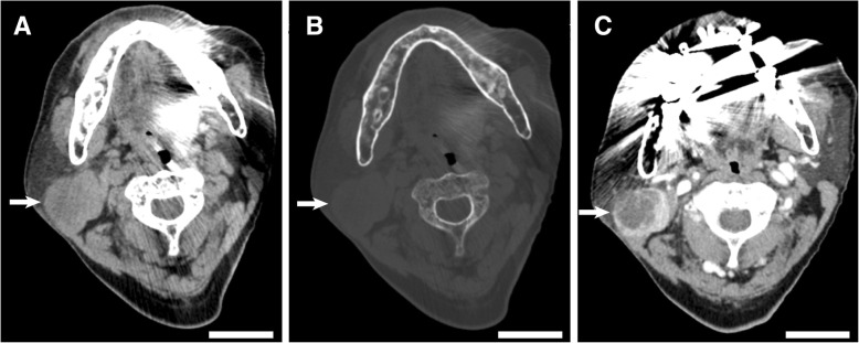
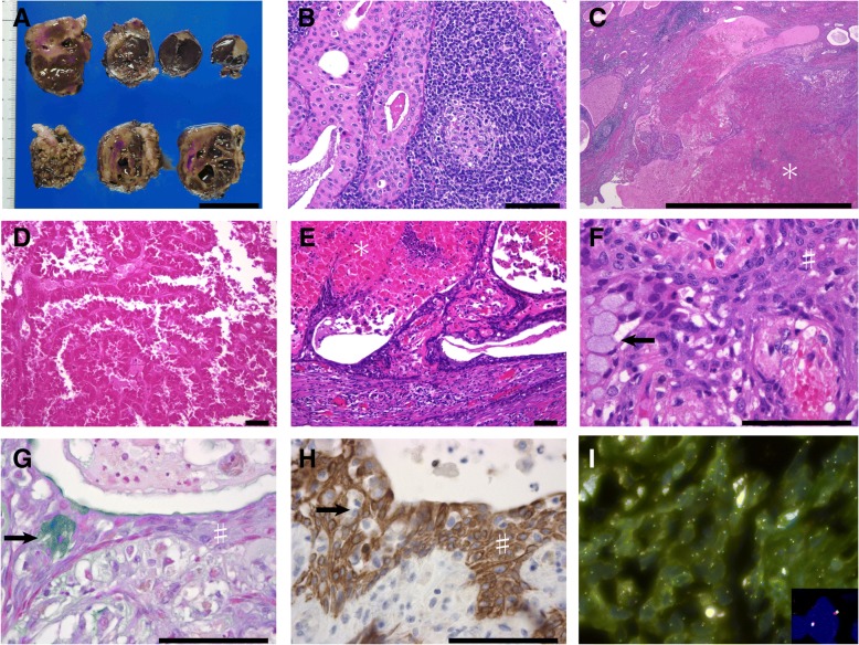
Case presentation: A 69-year-old Japanese man presented with a right parotid tumor. He had noticed the swelling on his right buccal region 1 year previously; the lesion had rapidly enlarged, with associated pain, 1 month previously. A radiological examination revealed a mass in the tail of the right parotid gland. Superficial parotidectomy was performed. On histological examination, the mass showed typical focal features of Warthin tumor; other areas showed coagulation necrosis of the tumor. These areas were surrounded by non-oncocytic epithelium comprising squamous and mucinous epithelial cells. Although cellular atypia of the non-oncocytic epithelium was not observed, a mixture of squamous and mucinous cells and lack of abundant lymphoid tissue mimicked low-grade mucoepidermoid carcinoma. Based on the results of fluorescence in situ hybridization, MAML2 gene rearrangement was not present in the typical portions of Warthin tumor and the mucoepidermoid carcinoma-like lesion. Therefore, a metaplastic or infarcted Warthin tumor was diagnosed. Our patient was disease-free 8 months after surgery.
Conclusions: Clinicians need to know that pain is a clinical symptom of infarcted/metaplastic Warthin tumor. Pathologists should be aware that a metaplastic Warthin tumor can mimic a low-grade mucoepidermoid carcinoma. Our case showed a mucoepidermoid carcinoma-like lesion that was confined near the area of tumor necrosis, and neither cytological atypia nor apparent invasive growth was present. These findings appeared to be histological clues of a metaplastic Warthin tumor rather than a mucoepidermoid carcinoma. Careful clinicopathological evaluation as well as genetic studies are needed to clarify the distinction between mucoepidermoid carcinoma and metaplastic portions of Warthin tumors.
Keywords: Metaplasia; Mucoepidermoid carcinoma; Necrotizing sialometaplasia; Warthin tumor.
Conflict of interest statement
Consent for publication
Written informed consent was obtained from the patient for publication of this case report and the use of accompanying images. A copy of the written consent is available for review by the Editor-in-Chief of this journal.
Competing interests
The authors declare that they have no competing interests.
Publisher’s Note
Springer Nature remains neutral with regard to jurisdictional claims in published maps and institutional affiliations.
Figures
References
-
- Nagao T, Gnepp DR, Simpson RHW, Vielh P. Warthin tumour. In: El-Naggar AK, JKC C, Grandis JR, Takata T, Slootweg PJ, editors. World Health Organization classification of head and neck tumors. Lyon: International Agency for Research on Cancer; 2017. pp. 188–189.
Publication types
MeSH terms
LinkOut - more resources
Full Text Sources
Medical
