. 2018 Dec 24:12:169-179.
doi: 10.2147/OTT.S184078. eCollection 2019.

LncRNA SNHG5 promotes growth and invasion in melanoma by regulating the miR-26a-5p/TRPC3 pathway

Affiliations

LncRNA SNHG5 promotes growth and invasion in melanoma by regulating the miR-26a-5p/TRPC3 pathway

Jun Gao et al. Onco Targets Ther. .

Abstract

Introduction: Melanoma has been reported as the most common malignancy in skin cancer. The small nucleolar RNA host gene 5 (SNHG5), an lncRNA, has been proven as a vital regulator in several types of carcinoma. This study was designed to investigate the detailed roles and possible mechanisms of SNHG5 in melanoma progression.

Methods: Quantitative real-time PCR (qRT-PCR) analysis was conducted to detect the expression levels of SNHG5, miR-26a-5p and transient receptor potential, canonical 3 (TRPC3) mRNA in melanoma tissues and cells. CCK-8 assay was used to measure the cell viability. Flow cytometry assays were performed to determine the cell cycle distribution and apoptosis. The invasive ability was assessed by a 24-well Transwell insert. Western blot analysis was employed to evaluate the protein expression of TRPC3. Dual luciferase reporter assay, RNA immunoprecipitation (RIP) assay, and RNA pull-down assay were applied to identify the interactions among SNHG5, miR-26a-5p and TRPC3.

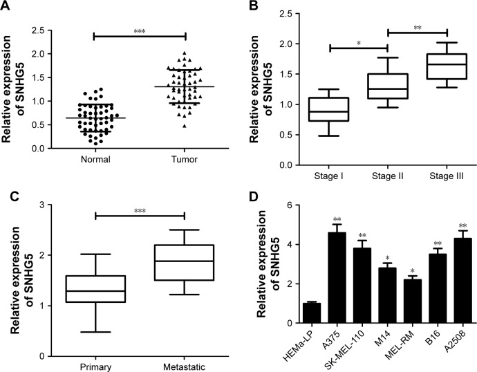
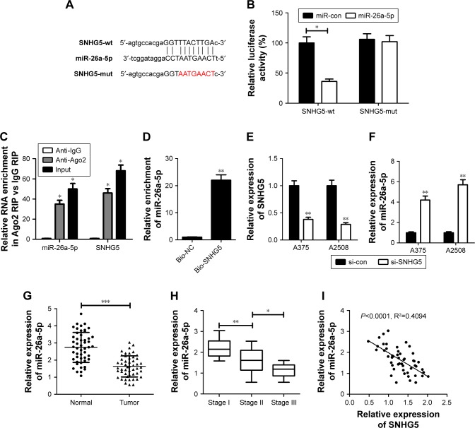
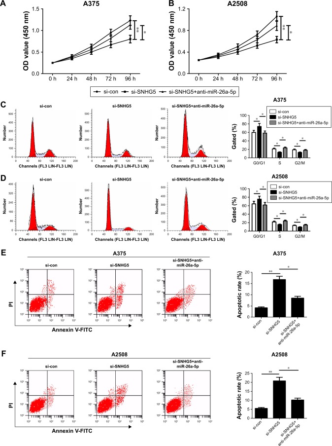
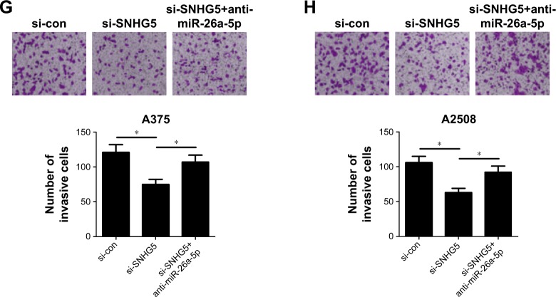
Results: The results showed that SNHG5 expression was increased in melanoma tumor tissues and cell lines. Higher SNHG5 expression was correlated with advanced pathogenic status. Moreover, SNHG5 could serve as a molecular sponge of miR-26a-5p. SNHG5 downregulation repressed proliferation, promoted apoptosis, and decreased invasion in melanoma cells, while these effects were greatly counteracted by miR-26a-5p inhibitor. Furthermore, miR-26a-5p directly targeted TRPC3 to suppress its expression, and this effect was aggravated following SNHG5 downregulation. Also, TRPC3 depletion exerted similar tumor-suppressive functions as SNHG5 knockdown.

Conclusion: SNHG5 promoted melanoma development by inhibiting miR-26a-5p and facilitating TRPC3 expression, highlighting the potential of SNHG5 as a novel target therapy for melanoma.

Keywords: SNHG5; TRPC3; cutaneum carcinoma; lncRNA; miR-26a-5p.


Conflict of interest statement

Disclosure The authors report no conflicts of interest in this work.

Figures

Figure 1
Figure 1
SNHG5 expression was increased in tumor tissues and cells of melanoma. Notes: qRT-PCR analysis of SNHG5 expression in tumor tissues and adjacent normal tissues of 50 melanoma patients (A), in different clinical pathological stages (B), in primary and metastatic melanoma tissues (C), as well as in melanoma cells (A375, SK-MEL-110, M14, MEL-RM, B16, and A2508) and normal human epidermal melanocytes (HEMa-LP) (D). *P<0.05, **P<0.01, ***P<0.001. Abbreviations: qRT, quantitative real-time; SNHG5, small nucleolar RNA host gene 5.
Figure 2
Figure 2
SNHG5 suppressed miR-26a-5p expression as a molecular sponge in melanoma cells. Notes: (A) The putative binding sites between SNHG5 and miR-26a-5p. (B) Dual luciferase reporter assays were conducted to detect the luciferase activity in A375 cells co-transfected with SNHG5-wt or SNHG5-mut reporter and miR-con or miR-26a-5p. (C) RIP assays were performed using Ago2 antibody or control IgG antibody in A375 cells, and then qRT-PCR analysis was used to measure the enrichment degrees of SNHG5 and miR-26a-5p in coprecipitated RNA. (D) RNA pull-down experiment was carried out to determine the binding between SNHG5 and miR-26a-5p in A375 cells. (E, F) qRT-PCR analysis of SNHG5 and miR-26a-5p expressions in A375 and A2508 cells after transfection with si-con or si-SNHG5. (G) MiR-26a-5p expression in tumor tissues and corresponding non-cancerous tissues of 50 melanoma patients. (H) MiR-26a-5p expression in tumor tissues at different clinical pathological stages. (I) The negative correlation between SNHG5 and miR-26a-5p expressions in 50 cases of melanoma tumor tissues. *P<0.05, **P<0.01, ***P<0.001. Bio-NC, a biotinylated lncRNA that is not complementary to miR-26a-5p was employed as a negative control. Abbreviations: mut, mutant; qRT, quantitative real-time; RIP, RNA immunoprecipitation; si-con, scrambled siRNAs; SNHG5, small nucleolar RNA host gene 5; WT, wild type.
Figure 3
Figure 3
SNHG5 knockdown suppressed proliferation, induced cell cycle arrest, promoted apoptosis, and decreased invasion in melanoma cells. Notes: A375 and A2508 cells were transfected with either si-SNHG5 alone, or together with anti-miR-26a-5p, followed by the examination of cell proliferation capability by CCK-8 (A, B), cell cycle distribution (C, D) and apoptosis (E, F) by flow cytometry analysis, as well as cell invasion ability by transwell invasion assay (G, H). *P<0.05, **P<0.01. Magnification ×200. Abbreviations: CCK-8, Cell Counting Kit-8; FITC, fluorescein isothiocyanate; OD, optical density; PI, propidium iodide; si-con, scrambled siRNAs; SNHG5, small nucleolar RNA host gene 5; RIP, RNA immunoprecipitation.
Figure 3
Figure 3
SNHG5 knockdown suppressed proliferation, induced cell cycle arrest, promoted apoptosis, and decreased invasion in melanoma cells. Notes: A375 and A2508 cells were transfected with either si-SNHG5 alone, or together with anti-miR-26a-5p, followed by the examination of cell proliferation capability by CCK-8 (A, B), cell cycle distribution (C, D) and apoptosis (E, F) by flow cytometry analysis, as well as cell invasion ability by transwell invasion assay (G, H). *P<0.05, **P<0.01. Magnification ×200. Abbreviations: CCK-8, Cell Counting Kit-8; FITC, fluorescein isothiocyanate; OD, optical density; PI, propidium iodide; si-con, scrambled siRNAs; SNHG5, small nucleolar RNA host gene 5; RIP, RNA immunoprecipitation.
Figure 4
Figure 4
The tumor-suppressive roles of TRPC3 silencing were mediated by SNHG5/miR-26a-5p. Notes: (A) The predicted complementary sequence between miR-26a-5p and TRPC3-3′UTR region. (B) Luciferase activity was evaluated in A375 cells co-transfected with TRPC3-wt or TRPC3-mut reporter and miR-con, miR-26a-5p, miR-26a-5p+vector, or miR-26a-5p+pcDNA-SNHG5. (C) Western blot analysis was performed in A375 and A2508 cells to assess the effect of miR-26a-5p overexpression or SNHG5 knockdown on TRPC3 expression. Every experiment was performed in triplicate and repeated three times. (D) TRPC3 expression in tumor tissues and adjacent normal tissues of 50 melanoma patients. (E) TRPC3 mRNA expression in tumor tissues at different clinical pathological stages. (F, G) The correlation assay between TRPC3 and miR-26a-5p or SNHG5 in 50 cases of melanoma tumor tissues. A375 cells were transfected with si-con or si-TRPC3, followed by qRT-PCR analysis of TRPC3 expression (H), MTT analysis of cell proliferation capability (I), flow cytometry assay of cell cycle distribution (J) and apoptotic rate (K), as well as transwell invasion assay of invasion ability (L). *P<0.05, **P<0.01, ***P<0.001. Abbreviations: FITC, fluorescein isothiocyanate; mut, mutant; miR-con, scrambled miRNA control; OD, optical density; qRT, quantitative real-time; si-con, scrambled siRNAs; SNHG5, small nucleolar RNA host gene 5; TRPC3, transient receptor potential, canonical 3; WT, wild type.
Figure 4
Figure 4
The tumor-suppressive roles of TRPC3 silencing were mediated by SNHG5/miR-26a-5p. Notes: (A) The predicted complementary sequence between miR-26a-5p and TRPC3-3′UTR region. (B) Luciferase activity was evaluated in A375 cells co-transfected with TRPC3-wt or TRPC3-mut reporter and miR-con, miR-26a-5p, miR-26a-5p+vector, or miR-26a-5p+pcDNA-SNHG5. (C) Western blot analysis was performed in A375 and A2508 cells to assess the effect of miR-26a-5p overexpression or SNHG5 knockdown on TRPC3 expression. Every experiment was performed in triplicate and repeated three times. (D) TRPC3 expression in tumor tissues and adjacent normal tissues of 50 melanoma patients. (E) TRPC3 mRNA expression in tumor tissues at different clinical pathological stages. (F, G) The correlation assay between TRPC3 and miR-26a-5p or SNHG5 in 50 cases of melanoma tumor tissues. A375 cells were transfected with si-con or si-TRPC3, followed by qRT-PCR analysis of TRPC3 expression (H), MTT analysis of cell proliferation capability (I), flow cytometry assay of cell cycle distribution (J) and apoptotic rate (K), as well as transwell invasion assay of invasion ability (L). *P<0.05, **P<0.01, ***P<0.001. Abbreviations: FITC, fluorescein isothiocyanate; mut, mutant; miR-con, scrambled miRNA control; OD, optical density; qRT, quantitative real-time; si-con, scrambled siRNAs; SNHG5, small nucleolar RNA host gene 5; TRPC3, transient receptor potential, canonical 3; WT, wild type.

References

    1. Siegel RL, Miller KD, Jemal A. Cancer statistics, 2016. CA Cancer J Clin. 2016;6666(1):7–30. - PubMed
    1. Siegel RL, Miller KD, Jemal A. Cancer Statistics, 2017. CA Cancer J Clin. 2017;6767(1):7–30. - PubMed
    1. Luke JJ, Schwartz GK. Chemotherapy in the management of advanced cutaneous malignant melanoma. Clin Dermatol. 2013;31(3):290–297. - PMC - PubMed
    1. Boada A, Carrera C, Segura S, et al. Cutaneous toxicities of new treatments for melanoma. Clin Transl Oncol. 2018;20(11):1373–1384. - PubMed
    1. Fang Y, Fullwood MJ. Roles, functions, and mechanisms of long non-coding RNAs in cancer. Genomics Proteomics Bioinformatics. 2016;14(1):42–54. - PMC - PubMed