. 2018 Aug 8;5(5):492-498.
doi: 10.1002/mdc3.12626. eCollection 2018 Sep-Oct.

Possible Post-Traumatic Focal Dystonia Associated with Tau Pathology Localized to Putamen-Globus Pallidus

Affiliations

Possible Post-Traumatic Focal Dystonia Associated with Tau Pathology Localized to Putamen-Globus Pallidus

Diego Iacono et al. Mov Disord Clin Pract. .

Abstract

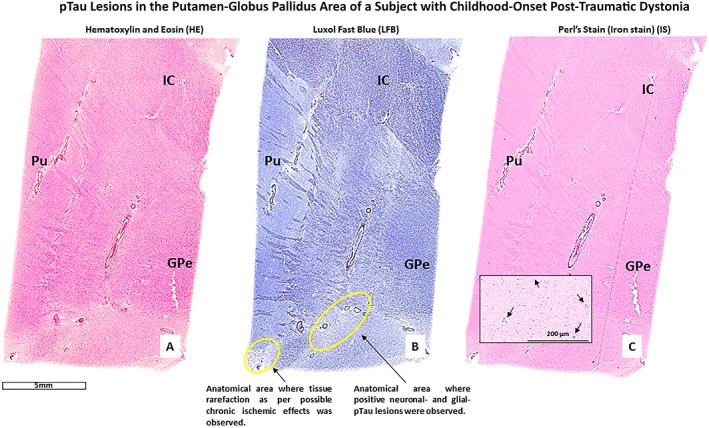
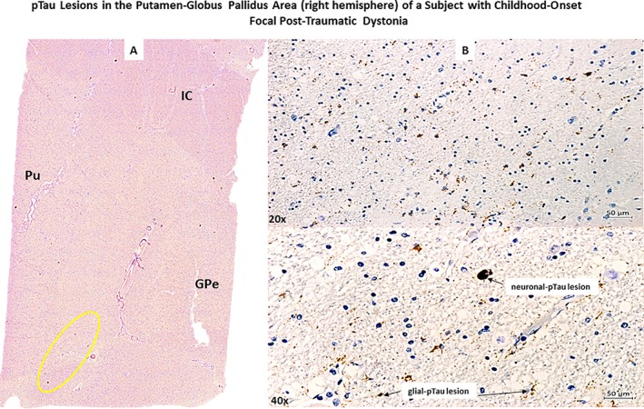
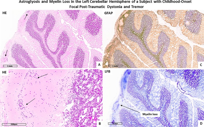
Background: Dystonia is often associated with damage to basal ganglia (BG), but neuropathological assessments of these cases are infrequent.

Methods: A brain was assessed with possible post-traumatic focal dystonia that appeared after an accident occurred during childhood.

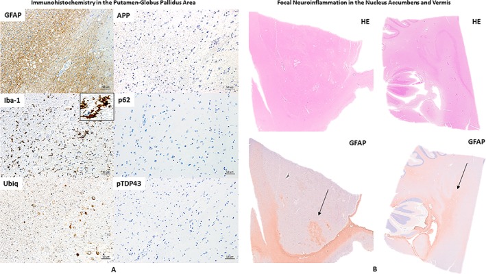
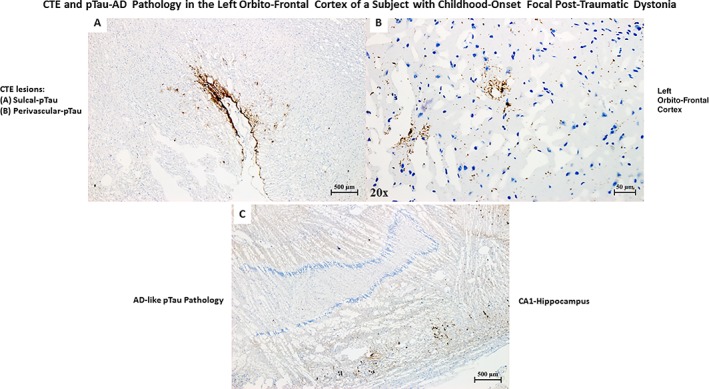
Results: Tau pathology was found within putamen and globus pallidus of the right hemisphere, and chronic traumatic encephalopathy (CTE) was observed in the cortex of the left hemisphere. No diffuse axonal injury (DAI), β-amyloid, ubiquitin, p62, or pTDP43 pathology was found.

Conclusions: Post-traumatic dystonia could be associated with post-traumatic tau pathology formation. However, more cases are necessary to establish causality. The tau lesions found in the BG of this patient did not fit within CTE criteria. We hypothesize that due to the anatomo-histological characteristics of the BG, tau pathology associated with brain traumas produce histopathological patterns different from sulcal-tau pathology, which is the only tau pathology distribution currently accepted as pathognomonic of CTE.

Keywords: CTE spectrum; basal ganglia; post‐traumatic dystonia; tau pathology; traumatic brain injury.


Figures

Figure 1
Figure 1
The figure shows histological sections from the right Putamen (Pu) ‐ Globus Pallidus external (GPe) area of a patient with possible child‐onset post‐traumatic focal dystonia. Part A, B, and C are images of three consecutive sections respectively stained with hematoxylin and Eosin (HE), Luxol Fast Blue (LFB), and iron stain (Perl's stain). Note: the presence of iron (hemosiderin) deposition (blue spots, part C) in the lenticular region of the right BG of this patient.
Figure 2
Figure 2
The figure (Part A) shows a section stained with AT8 antibody (anti‐phosphorylated‐tau antibody) of the right Putamen (Pu)‐Globus Pallidus external (GPe) area of a patient with possible child‐onset post‐traumatic focal dystonia. (Part B). Top and bottom images show neuronal‐ and glia‐tau lesions respectively at a higher progressive magnification (20x, 40x objectives).
Figure 3
Figure 3
(A) Consecutive histological sections respectively immunostained for GFAP, iba‐1 (to note the microglia activation as shown by the black arrow), Ubiq, APP (negative), p62 (negative), and pTDP43 (negative) showing the same anatomical area (right Putamen (Pu)‐Globus Pallidus external (GPe). (B) The nucleus accumbens and cerebellar cortex (including vermis region) of the right hemisphere stained with HE and GFAP. To note the distinct areas of focal neuroinflammation (GFAP positive) in both regions (black arrow).
Figure 4
Figure 4
(A) pTau pathology (AT8 stain) pathognomonic of chronic traumatic encephalopathy (CTE) in the orbitofrontal frontal cortex of the left hemisphere of a patient with a single TBI occurred during childhood. (B) Perivascular pTau pathology, part of CTE pathology. (C)pTau pathology (AT8 stain) in the CA1 region of the left hippocampus typical of Alzheimer's disease (AD) pathology. The patient was never diagnosed with memory or other cognitive deficits until death. Based on the age of the patient, the presence of pTau pathology in the hippocampus is compatible with a diagnosis primary age‐related tauopathy (PART).
Figure 5
Figure 5
The figure shows reactive astrogliosis as evidenced by hematoxylin and eosin (HE; A and B) and GFAP (C) stain. Panel D shows cerebellar myelin loss as evidenced by Luxol Fast Blue (LFB) stain.

Similar articles

Cited by

References

    1. Defazio G, Abbruzzese G, Livrea P, Berardelli A. Epidemiology of primary dystonia. Lancet Neurol. 2004;3:673–678. - PubMed
    1. Steeves TD, Day L, Dykeman J, Jette N, Pringsheim T. The prevalence of primary dystonia: a systematic review and meta‐analysis. Mov Disord. 2012;27:1789–1796. - PubMed
    1. Lohmann K, Klein C. Update on the Genetics of Dystonia. Curr Neurol Neurosci Rep. 2017;17:26. - PubMed
    1. Koller WC, Wong GF, Lang A. Posttraumatic movement disorders: a review. Mov Disord. 1989;4:20–36. - PubMed
    1. Jankovic J. Post‐traumatic movement disorders: central and peripheral mechanisms. Neurology. 1994;44:2006–2014. - PubMed

LinkOut - more resources