Review
. 2019 Apr;126(4):455-471.
doi: 10.1007/s00702-019-01969-2. Epub 2019 Jan 14.

Adenosine heteroreceptor complexes in the basal ganglia are implicated in Parkinson's disease and its treatment

Affiliations
Review

Adenosine heteroreceptor complexes in the basal ganglia are implicated in Parkinson's disease and its treatment

Dasiel O Borroto-Escuela et al. J Neural Transm (Vienna). 2019 Apr.

Abstract

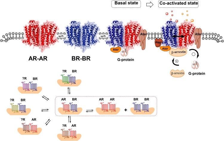
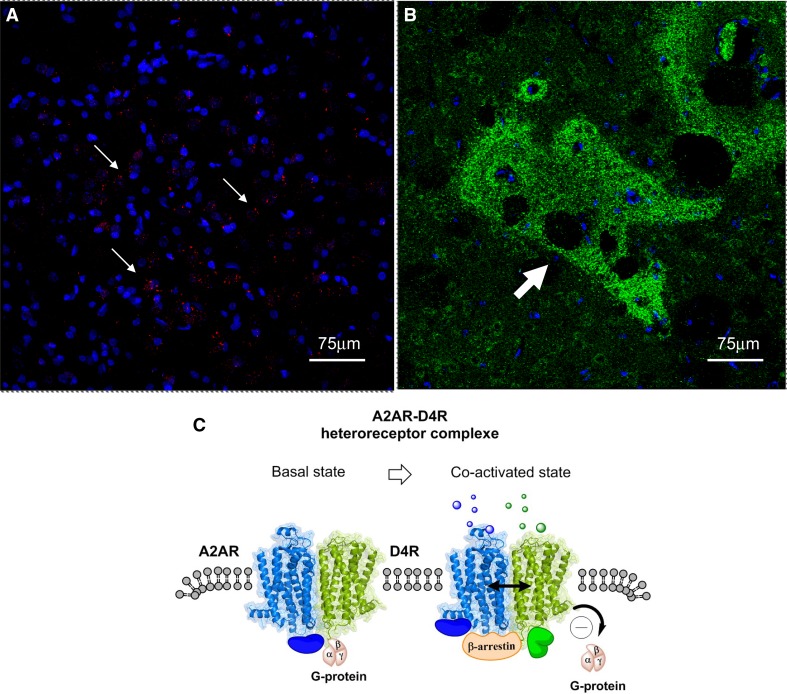
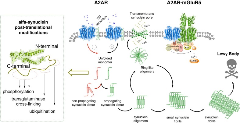
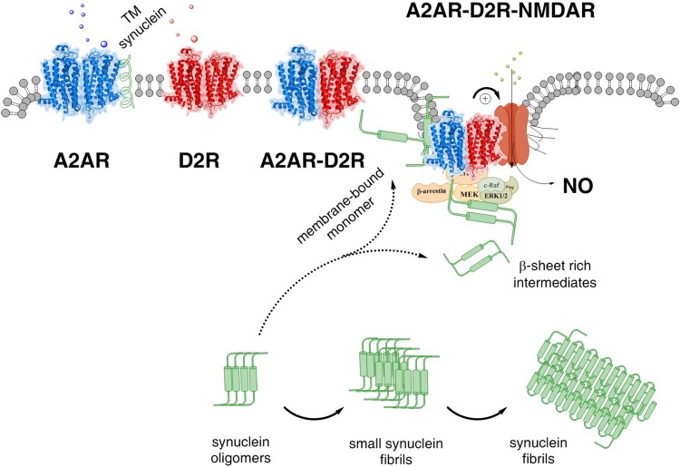
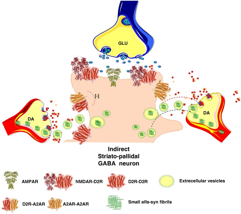
The adenosine homo, iso and heteroreceptor complexes in the basal ganglia play a highly significant role in modulating the indirect and direct pathways and the striosomal projections to the nigro-striatal DA system. The major adenosine receptor complexes in the striato-pallidal GABA neurons can be the A2AR-D2R and A2AR-D2R-mGluR5 receptor complexes, in which A2AR protomers and mGluR5 protomers can allosterically interact to inhibit D2R protomer signaling. Through a reorganization of these heteroreceptor complexes upon chronic dopaminergic treatment a pathological and prolonged inhibition of D2R receptor protomer signaling can develop with motor inhibition and wearing off of the therapeutic effects of levodopa and dopamine receptor agonists. The direct pathway is enriched in D1R in and around glutamate synapses enhancing the ability of these GABA neurons to be activated and increase motor initiation. The brake on these GABA neurons is in this case exerted by A1R forming A1R-D1R heteroreceptor complexes in which they allosterically inhibit D1R signaling and thereby reduce motor initiation. Upon chronic levodopa treatment a reorganization of the D1R heteroreceptor complexes develops with the formation of putative A1R-D1R-D3 in addition to D1R-D3R complexes in which D3R enhances D1R protomer signaling and may make the A1R protomer brake less effective. Alpha-synuclein monomers-dimers are postulated to form complexes with A2AR homo and heteroprotomers in the plasma membrane enhancing alpha-synuclein aggregation and toxicity. The alpha-synuclein fibrils formed in the A2AR enriched dendritic spines of the striato-pallidal GABA neurons may reach the surrounding DA terminals via extracellular-vesicle-mediated volume transmission involving internalization of the vesicles and their cargo (alpha-synuclein fibrils) into the vulnerable DA terminals, enhancing their degeneration followed by retrograde flow of these fibrils in the DA axons to the vulnerable nigral DA nerve cells.

Keywords: Adenosine receptor; Basal ganglia; G protein-coupled receptor; Heteroreceptor complexes; Neurodegeneration; Oligomerization; Parkinson’s diseases.


Figures

Fig. 1
Fig. 1
(Upper panel). Illustration of the fundamentals of the allosteric receptor–receptor interactions and the role of the receptor interface. A receptor heterodimer A–B is formed when certain amino acids in the two surfaces of the receptor interface of the heterodimer can bind to each other and form hot spots of considerable strength. Each heterodimer develops its own unique allosteric receptor–receptor interactions that must pass over the receptor interface, which can be located in transmembrane, intracellular and/or extracellular regions of the receptor. Upon, e.g., agonist activation of one receptor protomer the allosteric wave induced will pass over the receptor interface and induce either facilitatory or antagonistic allosteric changes in the function of the other receptor protomer. It can also involve a switch of, e.g, signaling over Gi/o to signaling via another G protein or beta-arrestin (bottom panel). A heterodimer A–B can also be in balance with other receptor complexes of different types (XA, XB) in a synaptic plasma membrane region or an extrasynaptic plasma membrane region. A disturbance of the balance among the receptor complexes can lead to dysfunction in transmission and to mental and neurological disorders
Fig. 2
Fig. 2
a The red PLA positive clusters of A2AR–D4R heteroreceptor complexes are enriched in a distinct region of the sampled field from the dorsal striatum. White arrows point to some of the red PLA positive clusters. Nuclei have a blue color. b Green D4R immunoreactivity is shown as densely packed green dots forming islands (see white arrow) (striosomes) in the dorsal striatum. c A2AR–D4R heterodimers in the plasma membrane in the basal state and in the co-activated state. Upon coactivation of the A2AR protomer (Blue) and the D4R protomer (green) the D4R induced increase in Gi/o protein signaling is probably inhibited by the antagonistic allosteric A2AR–D4R interaction. At the same time beta-arrestin can be recruited to the A2AR–D4R heterodimer and become part of the A2AR–D4R signaling. The blue and green solid spheres represent different types of adapter proteins bound to the heterodimer and upon coactivation a new adapter protein (in green) can be recruited to the heterodimer
Fig. 3
Fig. 3
Possible molecular mechanism by which alpha-synuclein monomers/oligomers/synuclein fibrils can modulate the A2AR homo-heteroreceptor complexes and their balance in the plasma membrane. In the left part it is proposed that monomeric alpha-synuclein transmembrane (TM) peptides can become linked to the A2AR homoreceptor complex and modulate the A2AR function. Under the modulation of the monomeric alpha-synuclein peptides the A2AR antagonist may favor the formation of non-propagating alpha-synuclein dimers (pathway highlighted in red). Instead the A2A receptor agonist induced A2AR activation (pathway highlighted in green) may in the alpha-synuclein-A2AR complex produce signals that favor the propagation of alpha-synuclein dimers/oligomers into small and large synuclein aggregates that accumulate in Lewy bodies.In the far left several mechanisms are illustrated that can produce posttranslational modifications of alpha-synuclein. It involves changes in phosphorylation, transglutaminase cross-linking and ubiquitination and may play a role in the transformation of alpha-synuclein into propagating alpha-synuclein dimers. Ring-like synuclein oligomers can also be formed, which enter the plasma membrane and there produce beta sheet structures that associate and produce pores through which calcium ions may pass. In the A2AR–mGluR5 heteromer, shown as the coming together of two homodimers (A2AR homodimer in blue and mGluR5 homodimer in green), the signaling pathways are illustrated. Changes in the activity of protein kinases like PKA, PKC and calcium–calmodulin kinase II can have a role in the modulation of the synuclein aggregation process
Fig. 4
Fig. 4
The A2AR–D2R–NMDAR complex and its balance with A2AR–A2AR and D2R–D2R homodimers and A2AR–D2R heterdimers are illustrated. The alpha-synuclein monomer (alpha conformation) may also bind to the A2AR protomer of the A2AR–D2R–NMDAR complex, enhancing A2AR protomer activation and its allosteric inhibition of the D2R signaling. As a result the allosteric inhibition by the D2R protomer of the NMDAR protomer is reduced and NMDAR function may become substantially enhanced with increased calcium influx through its ion channels (see + sign) leading to a coupling to nitric oxide (NO) production and toxicity. Beta sheet rich intermediates of alpha-synuclein peptides are proposed to bind to the intracellular loops and C terminal of the receptor protomers of this heteroreceptor trimeric complex and modulate their signaling. In addition, the signaling of the G proteins and beta-arrestin as well of other associated proteins like kinases in the signaling pathways may become modulated
Fig. 5
Fig. 5
Illustration of how A2AR signaling can enhance the formation alpha-synuclein fibrils in the dendritic spines of the striato-pallidal GABA neurons. It is also indicated how the alpha synuclein fibrils can be transported from the dendrites of the dorsal striato-pallidal GABA neurons to the DA nerve terminals of the vulnerable nigrostriatal DA neurons involving extracellular vesicle mediated volume transmission. The fibrils can exist in exosomes and upon release internalized into the surrounding striatal DA nerve terminals followed by retrograde transport in the DA axons to the nigral DA cell bodies. This hypothesis is inline with the view that neurodegeneration of the nigro-striatal DA neurons starts in the DA nerve terminal networks

References

    1. Agnati LF, Fuxe K. Extracellular-vesicle type of volume transmission and tunnelling-nanotube type of wiring transmission add a new dimension to brain neuro-glial networks Philosophical transactions of the Royal Society of London Series B. Biol Sci. 2014 - PMC - PubMed
    1. Anden NE, Carlsson A, Dahlstroem A, Fuxe K, Hillarp NA, Larsson K. Demonstration and mapping out of nigro-neoestriatal dopamine neurons. Life Sci. 1964;3:523–530. - PubMed
    1. Antonelli T, Fuxe K, Agnati L, Mazzoni E, Tanganelli S, Tomasini MC, Ferraro L. Experimental studies and theoretical aspects on A2A/D2 receptor interactions in a model of Parkinson’s disease. relevance for L-dopa induced dyskinesias. J Neurol Sci. 2006;248:16–22. - PubMed
    1. Audet N, Galés C, Archer-Lahlou E, Vallières M, Schiller PW, Bouvier M, Pineyro G. Bioluminescence resonance energy transfer assays reveal ligand-specific conformational changes within preformed signaling complexes containing delta-opioid receptors and heterotrimeric G proteins. J Biol Chem. 2008;283:15078–15088. - PMC - PubMed
    1. Azdad K, Gall D, Woods AS, Ledent C, Ferre S, Schiffmann SN. Dopamine D2 and adenosine A2A receptors regulate NMDA-mediated excitation in accumbens neurons through A2A-D2 receptor heteromerization Neuropsychopharmacology: official publication of the American College. Neuropsychopharmacology. 2009;34:972–986. - PMC - PubMed

Publication types

Substances