

Recent Advances in Imaging of Hypertensive Heart Disease
- PMID: 30637533
- PMCID: PMC6400461
- DOI: 10.1007/s11906-019-0910-6
Recent Advances in Imaging of Hypertensive Heart Disease
Abstract
Purpose of review: To review recent advances in the imaging of hypertensive heart disease (HHD) with an emphasis on developments in the imaging of diffuse myocardial fibrosis using cardiac magnetic resonance (CMR).
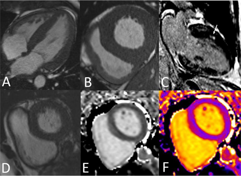
Recent findings: HHD results from long-standing hypertension and is characterized by the development of left ventricular hypertrophy and diffuse interstitial fibrosis. Diffuse fibrosis traditionally required endomyocardial biopsy to diagnose, but recent developments using T1 mapping in CMR allow for noninvasive assessment. Studies using T1 mapping have shown an increase in extracellular volume fraction (ECV) in patients with HHD compared to normal controls, suggesting ECV can be used as a noninvasive marker for fibrosis in HHD. In addition to T1 mapping, other recent advances in HHD imaging include improvements in three-dimensional echocardiography, allowing for accurate real-time volumetric measurements, and the use of speckle tracking echocardiography to detect subclinical systolic dysfunction. Measurement of ECV using T1 mapping in CMR can be used as a noninvasive marker of diffuse myocardial fibrosis in HHD. While further studies are needed to validate this approach with larger patient cohorts, ECV can potentially be used to both monitor disease progression and assess therapeutic interventions in HHD.
Keywords: Cardiac magnetic resonance; Hypertensive heart disease; Speckle tracking echocardiography; T1 mapping; Three-dimensional echocardiography.
Figures
References
-
- Mozaffarian D, Benjamin EJ, Go AS, Arnett DK, Blaha MJ, Cushman M, et al. Heart Disease and Stroke Statistics—2016 Update. Circulation 2016;133. - PubMed
-
- Díez J, González A, López B, Querejeta R. Mechanisms of disease: pathologic structural remodeling is more than adaptive hypertrophy in hypertensive heart disease. Nat Clin Pract Cardiovasc Med 2005;2:209–16. - PubMed
-
- Georgiopoulou VV, Kalogeropoulos AP, Raggi P, Butler J. Prevention, Diagnosis, and Treatment of Hypertensive Heart Disease. Cardiol Clin 2010;28:675–91. - PubMed
-
- Levy D, Garrison RJ, Savage DD, Kannel WB, Castelli WP. Prognostic Implications of Echocardiographically Determined Left Ventricular Mass in the Framingham Heart Study. N Engl J Med 1990;322:1561–6. - PubMed
-
- Schillaci G, Verdecchia P, Porcellati C, Cuccurullo O, Cosco C, Perticone F. Continuous relation between left ventricular mass and cardiovascular risk in essential hypertension. Hypertens 2000;35:580–6. - PubMed
Publication types
MeSH terms
Grants and funding
LinkOut - more resources
Full Text Sources
Medical
Research Materials
