. 2019:615:373-406.
doi: 10.1016/bs.mie.2018.08.023. Epub 2018 Sep 18.

DNP-Assisted NMR Investigation of Proteins at Endogenous Levels in Cellular Milieu

Affiliations

DNP-Assisted NMR Investigation of Proteins at Endogenous Levels in Cellular Milieu

Whitney N Costello et al. Methods Enzymol. 2019.

Abstract

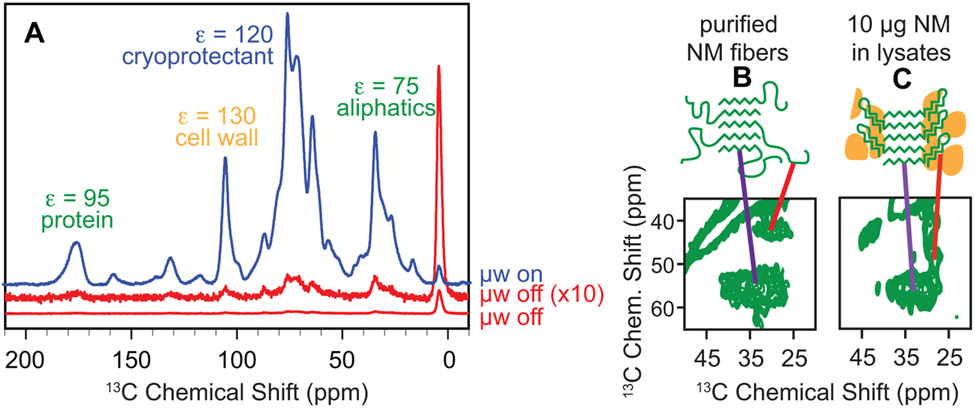
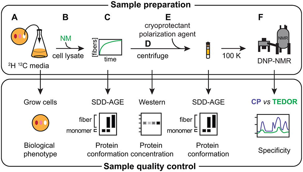
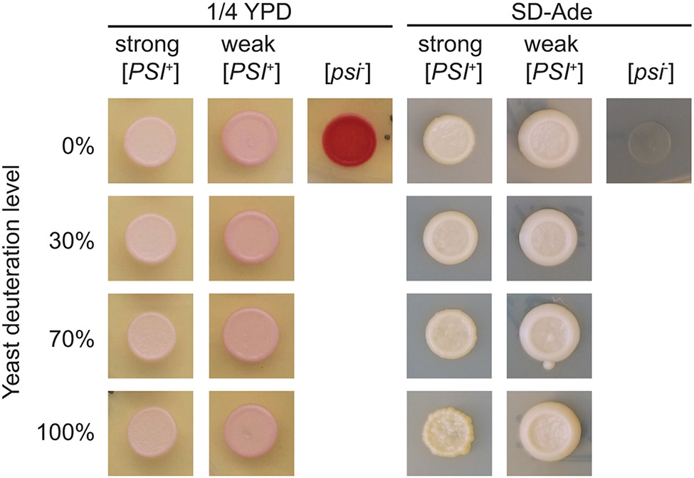
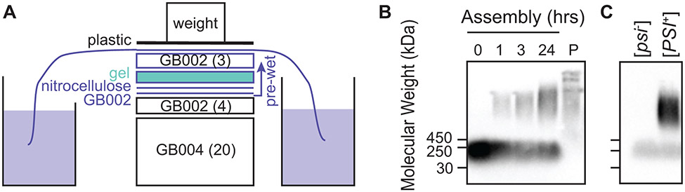
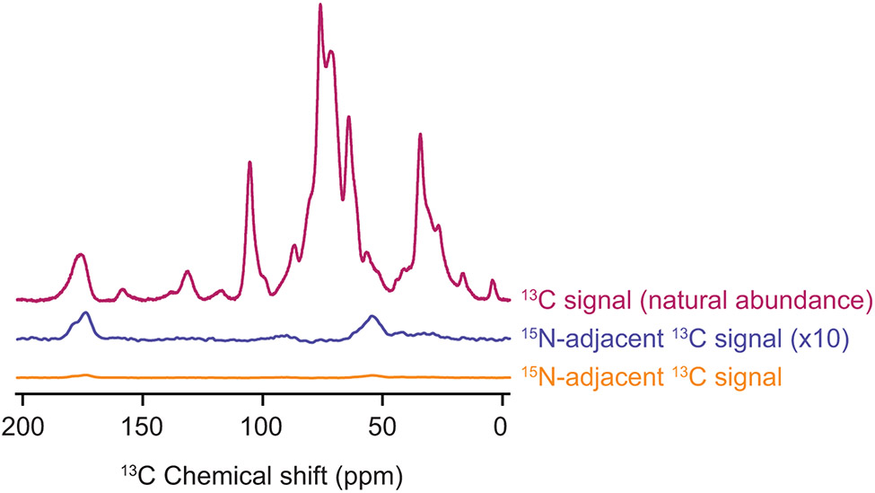
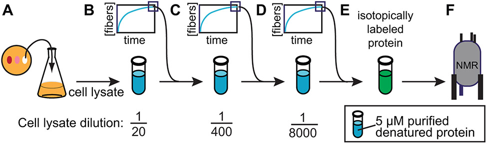
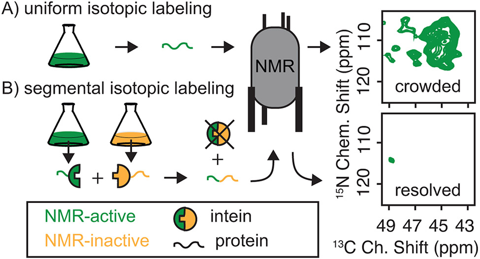
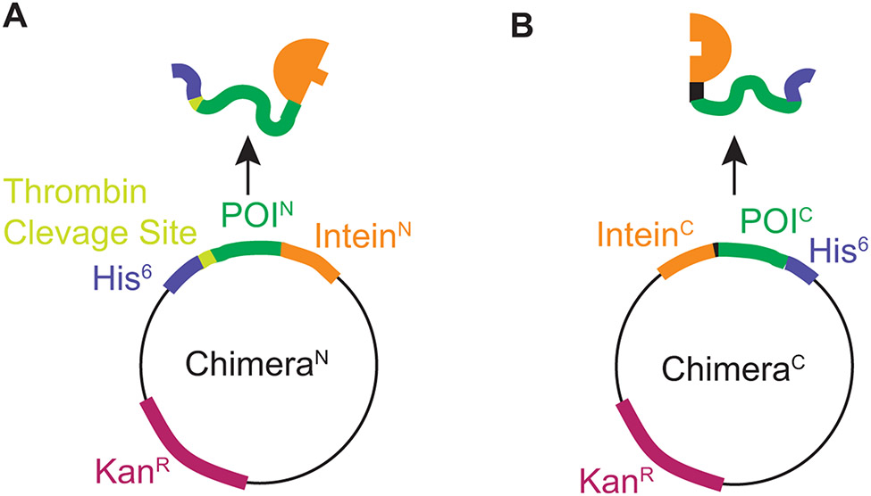
Structural investigations of biomolecules are typically confined to in vitro systems under extremely limited conditions. These investigations yield invaluable insights, but such experiments cannot capture important structural features imposed by cellular environments. Structural studies of proteins in their native contexts are not only possible using state-of-the-art sensitivity-enhanced (dynamic nuclear polarization, DNP) solid-state nuclear magnetic resonance (NMR) techniques, but these studies also demonstrate that the cellular context can and does have a dramatic influence on protein structure. In this chapter, we describe methods to prepare samples of isotopically labeled proteins at endogenous levels in cellular contexts alongside quality control methods to ensure that such samples accurately model important features of the cellular environment.

Keywords: Amyloids; Dynamic nuclear polarization; In situ structural biology; In-cell NMR; NMR; Prions; Sup35.


Figures

Figure 1:
Figure 1:
Dynamic nuclear polarization enhances NMR signals in cellular environments. A) One dimensional 13C{1H} CP spectra both with (blue) and without DNP enhancement (red). DNP NMR results in large signal enhancements (ε) for samples of per-deuterated cellular lysates in a 60:30:10 mixtures of d8-glycerol:D2O:H2O and 10 mM AMUPol at 600 MHz/385 GHz with ω/2π = 12.5 kHz and a sample temperature of 100 K. Spectroscopic selection for the small amount of uniformly isotopically labeled exogenous prepared protein that was added to the cellular lysate mixture reveals that the amyloid cores of this protein have similar conformations (purple line), the intrinsically disordered region (red line) has a very different chemical environment in (B) purified samples than it does in (C) in biological environments.
Figure 2:
Figure 2:
Sample preparation of proteins at endogenous levels in cellular environments for analysis by DNP NMR requires control of sample integrity. A) Yeast grown in per-deuterated media are phenotyped to ensure that changing the isotopic composition does not affect the biological phenotype of the cells. B) Exogenously-prepared isotopically enriched protein (e.g. NM) is added to lysed cells. C) The exogenously-added protein polymerizes using the cellular material as a template. Assembly of the added protein into the prion form in lysates is monitored by SDD-AGE. D) The insoluble portion of the sample is collected by centrifugation. Western blot analysis using an antibody specific for the protein of interest controls for protein degradation products and allows an estimate of the concentration of the added protein in the insoluble fraction. E) The lysate sample is prepared for DNP with the addition of cryoprotectants and the DNP polarization agent, like AMUPol, and frozen. Maintenance of the protein in the prion form through this process is also monitored by SDD-AGE. F) The specificity of the isotopic labeling schemes, particularly with respect to signals arising from natural abundance isotopes in the cellular lysate (see Table 1), are controlled for by comparison of the spectra of all 13C atoms versus only 13C that are within bonding distance of another isotopically enriched atom.
Figure 3:
Figure 3:
The yeast prion phenotypes are maintained during growth in media deuterated up to ~100%. A small volume (~ 5 μL) of yeast grown in media with a variety of deuteration levels is spotted on plates for phenotyping immediately before the cells are collected. A) The colony color phenotype on ¼ YPD is maintained for strong [PSI+] (white), weak [PSI+] (pink), and [psi] (red) yeasts cultured in media with a variety of deuteration levels. B) The growth phenotype on SD-ade is likewise maintained for strong [PSI+] (present), weak [PSI+] (present), and [psi] (absent) yeasts cultured in media with a variety of deuteration levels, indicating that the protein conformation responsible for this trait is maintained under these conditions.
Figure 4:
Figure 4:
Semi-Denaturing Detergent-Agarose Gel Electrophoresis (SDD-AGE) is used to visualize the aggregation state of recombinantly-expressed, exogenously-added proteins. After separation of large protein aggregates on agarose gels, the proteins are transferred to a nitrocellulose membrane using capillary transfer. Because the read-out is antibody based, SDD-AGE is appropriate for analysis of proteins at low concentrations in complex mixtures. A) Transfer of proteins to the nitrocellulose membrane is accomplished by capillary transfer. The agarose gel is placed in direct contact with a wet nitrocellulose membrane. The gel and membrane are placed on a stack of dry blotting paper, covered by wet blotting paper kept moist through the action of a wick in a reservoir of buffer. B) The kinetics of assembly of exogenously-added NM (visualized in this case by an antibody against a his6 tag) into the amyloid form in yeast lysates (e.g. Figure 2C). Monomeric NM is removed by centrifugation as evidenced by analysis of the pellet (P) as described in Figure 2D. C) Amyloid aggregates remain intact after the addition of cryoprotectants, polarization agents and freezing (e.g. Figure 2E). Moreover, these manipulations do not induce the formation of amyloid as evidenced by the lack of high molecular weight aggregates in samples prepared using [psi] yeast.
Figure 5:
Figure 5:
The combination of isotopic depletion of the cellular lysates with spectroscopic selection for adjacent isotopically labeled sites enables highly specific detection of proteins at endogenous concentrations. One dimensional 13C{1H} CP spectra report on all of the 13C in the sample (magenta). The TEDOR experiment with a 1.6 ms mixing time (orange) is selective for 13C sites with adjacent 15N atoms. Such sites are much less common (see Table 1) than 13C sites overall. The TEDOR spectra (orange) is multiplied by a factor of 4 to account for differences in experimental efficiency between the 13C CP experiment (magenta) and the TEDOR experiment (orange). It is further multiplied by a factor of 10 (blue) to allow comparison of the difference in features of the peak that reports on the carbonyl carbon chemical shift around 175 ppm. The peak near 175 ppm in the TEDOR spectra is shifted towards values that are consistent with beta sheet conformations and has a shoulder near 180 ppm, in line with the amino acid composition of the protein. Specificity for the 1 μM concentration of added protein from the cellular background can be obtained for samples of per-deuterated cellular lysates in a 60:30:10 mixtures of d8-glycerol:D2O:H2O and 10 mM AMUPol at 600 MHz/385 GHz with ω/2π = 12.5 kHz and a sample temperature of 100 K.
Figure 6:
Figure 6:
Fiber formation can be templated by yeast cell lysates. A) Cultured yeast cells harboring the desired prion phenotype are grown to mid-log phase and then lysed by bead beating. B) Lysates are diluted into buffer containing low concentrations of purified recombinant denatured protein. C & D) Subsequent rounds of polymerization are seeded by addition of a small volume of the previous reactions, effectively removing cellular material by dilution. E) These lysate templated seeds are used to polymerize a large volume of purified recombinant isotopically enriched protein into the amyloid form. F) This sample serves as a control for the effects of cellular environment on protein structure to complement study of the same protein in cellular lysates.
Figure 7:
Figure 7:
Segmental isotopic labeling simplifies NMR spectra. A) The glycine region of a 13C-15N correlation spectrum for uniformly 13C and 15N labeled NM has many peaks, consistent with a protein that contains 23 glycine resides. B) The same region for a full-length NM protein that is segmentally isotopically labeled has one peak, consistent with a single glycine in the isotopically labeled region. NMR was segmentally isotopically labeled by intein-mediated ligation of NM-intein chimeras purified from bacteria grown in media containing either NMR active (13C & 15N, green) or NMR inactive (12C & 14N, yellow) carbon and nitrogen sources.
Figure 8:
Figure 8:
Plasmid constructs designed to express chimeric proteins designed to produce segmentally isotopically labeled yeast prion protein. A) The plasmid for ChimeraN. The N terminal segment of the protein of interest is expressed as a chimera with a cleavable his6 tag for purification adjacent to the segment to be isotopically labeled and followed by the N terminal piece of the CfaGEP intein. B) The plasmid for ChimeraC. The C terminal segment of the protein of interest is expressed as a chimera that starts with the C terminal piece of the CfaGEP intein followed by the C terminal region of the protein of interest with a non-cleavable his6 tag. The intein has sequence requirements at the amino acids directly adjacent to the intein thus for optimal ligation, the intein introduces a cysteine mutation at the junction between the labeled and unlabeled region and the ligation site was chosen to be at a position the fulfilled the preference for a native bulky hydrophobic amino acid (Y) in the second position from the C-terminal intein ligation site.
Figure 9:
Figure 9:
Chimeric protein purification, chimeric protein ligation and spliced product purification can by analyzed be SDS-PAGE. Inteins are represented by half circles, exteins are represented by curved lines and isotopic enrichment and depletion is represented by green and yellow color, respectively. A) Chimeric proteins were purified to homogeneity using Ni-NTA resin. B) Ligation of ChimeraN and ChimeraC to form the full-length protein of interest was followed over 16 hours. Appearance of the full-length protein of interest and the N-terminal fragment of the intein serves as a criteria to assess the progression of the ligation reaction. The smaller (4 kDa) C-terminal intein fragment is often difficult to visualize by SDS-PAGE. C) The full-length protein, which contains two his6 tags, is purified away from the precursors by taking advantage of its increases affinity for Ni-NTA using a step gradient of increasing imidazole concentrations.
Figure 10:
Figure 10:
Custom tool for packing samples for DNP NMR analysis. A) An 18 gauge needle is flattened on one end to create a scoop to transfer ultracentrifuged cell pellets from the centrifuge tube to the NMR rotor using a B) ball-peen hammer.

References

    1. Abragam A (1983). The principles of nuclear magnetism. Clarendon Press.
    1. Akbey Ü, Franks WT, Linden A, Lange S, Griffin RG, van Rossum B-J, & Oschkinat H (2010). Dynamic nuclear polarization of deuterated proteins. Angewandte Chemie International Edition, 49(42), 7803–7806. 10.1002/anie.201002044 - DOI - PMC - PubMed
    1. Allen KD, Wegrzyn RD, Chernova TA, Müller S, Newnam GP, Winslett PA, et al. (2005). Hsp70 chaperones as modulators of prion life cycle: novel effects of Ssa and Ssb on the Saccharomyces cerevisiae prion [PSI+]. Genetics, 169(3), 1227–1242. 10.1534/genetics.104.037168 - DOI - PMC - PubMed
    1. Banci L, Barbieri L, Luchinat E, & Secci E (2013). Visualization of redox-controlled protein fold in living cells, 20(6), 747–752. 10.1016/j.chembiol.2013.05.007 - DOI - PubMed
    1. Bayro MJ, Debelouchina GT, Eddy MT, Birkett NR, MacPhee CE, Rosay M, et al. (2011). Intermolecular Structure Determination of Amyloid Fibrils with Magic-Angle Spinning and Dynamic Nuclear Polarization NMR. Journal of the American Chemical Society, 133(35), 13967–13974. 10.1021/ja203756x - DOI - PMC - PubMed

Publication types

LinkOut - more resources