. 2019 Feb 5;116(3):540-550.
doi: 10.1016/j.bpj.2018.12.009. Epub 2018 Dec 18.

Rickettsia Sca2 Recruits Two Actin Subunits for Nucleation but Lacks WH2 Domains

Affiliations

Rickettsia Sca2 Recruits Two Actin Subunits for Nucleation but Lacks WH2 Domains

Saif S Alqassim et al. Biophys J. .

Abstract

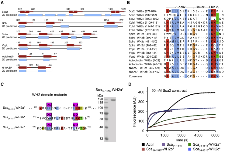
The Rickettsia ∼1800-amino-acid autotransporter protein surface cell antigen 2 (Sca2) promotes actin polymerization on the surface of the bacterium to drive its movement using an actin comet-tail mechanism. Sca2 mimics eukaryotic formins in that it promotes both actin filament nucleation and elongation and competes with capping protein to generate filaments that are long and unbranched. However, despite these functional similarities, Sca2 is structurally unrelated to eukaryotic formins and achieves these functions through an entirely different mechanism. Thus, while formins are dimeric, Sca2 functions as a monomer. However, Sca2 displays intramolecular interactions and functional cooperativity between its N- and C-terminal domains that are crucial for actin nucleation and elongation. Here, we map the interaction of N- and C- terminal fragments of Sca2 and their contribution to actin binding and nucleation. We find that both the N- and C-terminal regions of Sca2 interact with actin monomers but only weakly, whereas the full-length protein binds two actin monomers with high affinity. Moreover, deletions at both ends of the N- and C-terminal regions disrupt their ability to interact with each other, suggesting that they form a contiguous ring-like structure that wraps around two actin subunits, analogous to the formin homology-2 domain. The discovery of Sca2 as an actin nucleator followed the identification of what appeared to be a repeat of three Wiskott-Aldrich syndrome homology 2 (WH2) domains in the middle of the molecule, consistent with the presence of WH2 domains in most actin nucleators. However, we show here that contrary to previous assumptions, Sca2 does not contain WH2 domains. Instead, our analysis indicates that the region containing the putative WH2 domains is folded as a globular domain that cooperates with other parts of the Sca2 molecule for actin binding and nucleation.


Figures

Figure 1
Figure 1
Intramolecular interaction of the N- and C-terminal regions of Sca2. (A) Domain organization of Sca2 and constructs used in this study (SS, signaling sequence; NRD, N-terminal repeat domain; PRD1,2, proline-rich domains; WH2a–c, putative WH2 domains; CRD, C-terminal repeat domain; AC, predicted autochaperone domain; TD, translocator domain). (B) ITC titration of 200 μM Sca34–670 (in the syringe) into 20 μM Sca868–1515 (in the cell). The experiment was performed at 25°C. The dissociation constant (KD) and binding stoichiometry (N) derived from fitting of the binding isotherm are listed. Errors correspond to the SD of the fits. Open symbols correspond to titrations into buffer. (CH) Analytical size exclusion chromatography (SEC) and SDS-PAGE analyzes of mixtures of untagged N- and C-terminal Sca2 constructs as indicated. The gels shown as insets correspond to the SEC fractions of the peak, indicated by a dashed box. (C) shows that the premixed constructs Sca34–670 and Sca868–1515 run together by SEC, consistent with the formation of a complex. (D)–(F) show that the N-terminal fragment Sca34–670 runs separately from N- and C-terminal deletions of construct Sca868–1515 when premixed, consistent with lack of interaction. (G) and (H) show that the C-terminal fragment Sca868–1515 runs separately from N- and C-terminal deletions of constructs Sca34–670 when premixed, consistent with lack of interaction. The SEC traces of the Sca2 constructs are color-coded as indicated. To see this figure in color, go online.
Figure 2
Figure 2
Interaction of Sca2 constructs with monomeric actin. (A) ITC titration of 89 μM LatB-actin (in the syringe) into 8.3 μM Sca34–1515 (in the cell). The experiment was performed at 20°C. A two-site binding model produced a better fit of the data to a binding isotherm than a one-site binding model, as indicated by the χ2 values and realistic stoichiometry. The χ2 values reported here are reduced, i.e., divided by degrees of freedom. Note that although the χ2 value clearly defines the best fitting model for a particular titration, they are not generally comparable across different titrations because the data are not weighted and the χ2 depends on the magnitude of the scale. The dissociation constant (KD) and binding stoichiometry (N) derived from the fits are listed. Errors correspond to the SD of the fits. (B) ITC titration of 220 μM Sca34–670 (in the syringe) into 22 μM LatB-actin (in the cell). The experiment was performed at 12.7°C. A two-site binding model produced an almost equally good fit of the data to a binding isotherm than a one-site binding model, but is prefered based on a more realistic stoichiometry of the interaction. A two-site binding model is also supported by the fact that this fragment of Sca2 has nucleation activity (12). The dissociation constant (KD) and binding stoichiometry (N) derived from the fits are listed. Errors correspond to the SD of the fits. (C) ITC titration of 444 μM Sca869–1060 (in the syringe) into 16.6 μM LatB-actin (in the cell). The experiment was performed at 20°C. The dissociation constant (KD) and binding stoichiometry (N) derived from fitting of the binding isotherm are listed. Errors correspond to the SD of the fits. (D) ITC titration of 396 μM Sca1090–1355 (in the syringe) into 19 μM LatB-actin (in the cell). The experiment was performed at 20°C. No binding is reported because the experiment could not be fitted to a binding isotherm. The different fits are color-coded as indicated. To see this figure in color, go online.
Figure 3
Figure 3
The region 868–1023 of Sca2 is critical for nucleation but lacks WH2 domains. (A) A cartoon representation of the WH2-domain-containing regions of various cytoskeletal proteins containing tandem repeats of WH2 domains. WH2 domains are colored red, with a cylinder representing the α-helix of the domain. Secondary structure predictions obtained with the program Jpred (45) are also shown for each protein (two-dimensional prediction), with blue cylinders representing predicted α-helices. (B) Alignment of the sequences of the WH2 domains represented in (A), showing the regions corresponding to the N-terminal α-helix and LKKV motif, as well as the consensus sequence at the bottom. (C) Representation of the three mutants of Sca34–1515 (full-length passenger domain) targeting the putative WH2 domains. Each mutant carries four aspartic acid mutations of highly conserved residues of the canonical sequence of the domain. The SDS-PAGE shown on the right illustrates the purity of one of the mutants (Sca34–1515-WH2a). (D) A time course of the polymerization of 2 μM Mg-ATP-actin (6% pyrene labeled) alone and with the addition of 50 nM Sca34–1515 wild type and WH2 domain mutants depicted in (C) (color-coded as indicated). To see this figure in color, go online.
Figure 4
Figure 4
The putative WH2 domains of Sca2 do not bind actin. (A) A representation of four WH2 domain constructs expressed here as fusion proteins with maltose-binding protein (MBP). A 15-amino-acid flexible linker separates the MBP moiety at the N-terminus from the WH2 domain of WAVE1 (control) or the three putative WH2 domains of Sca2. (B) Purity of proteins illustrated in (A) assessed by SDS-PAGE analysis. (C) ITC titration of 254 μM WAVE1 WH2a into 18.5 μM LatB-actin. The experiment was performed at 25°C. The dissociation constant (KD) and binding stoichiometry (N) derived from fitting of the binding isotherm are listed. Errors correspond to the SD of the fits. (DF) ITC titrations of the three constructs of putative WH2 domains of Sca2 into LatB-actin. The concentrations of the proteins in the syringe and in the cell are indicated. The titrations could not be fitted to binding isotherms because none of the putative WH2 domains of Sca2 appeared to bind LatB-actin. To see this figure in color, go online.
Figure 5
Figure 5
Proposed mechanism of nucleation and elongation by Rickettsia Sca2. By analogy with eukaryotic formins, Sca2 is proposed to form a ring-like structure around the barbed end of the actin filament, contacting primarily two actin subunits and possibly a third incoming subunit. The data presented here support this model and suggest that the N- and C-terminal regions of Sca2 (Sca34–670 and Sca868–1515) behave in a way analogous to the FH2 domain of formins. Thus, we have shown here that these two fragments bind actin individually but with weak affinity, whereas the full-length passenger domain binds two actin subunits with high affinity and has both nucleation and elongation activities, although the individual fragments are mostly inactive (11, 12). Furthermore, deletions at either end of these two fragments disrupt their ability to interact with each other, suggesting that they interact head to tail to form a ring-like structure analogous to the formin FH2 domain. We have also shown that Sca2 does not contain WH2 domains. The region previously thought to contain the repeat of three WH2 domains (Sca869–1060) binds actin as a whole but only one actin subunit and in manner clearly distinct from that of canonical WH2 domains. Accordingly, this region is represented here as forming part of the ring-like structure of Sca2 and is renamed as the “middle domain” because of its location in the sequence. The two proline-rich domains, implicated in the recruitment of profilin-actin for barbed end elongation (12), may project out because they form part of loops that are predicted to be unstructured. To see this figure in color, go online.

Similar articles

Cited by

References

    1. Bugalhão J.N., Mota L.J., Franco I.S. Bacterial nucleators: actin’ on actin. Pathog. Dis. 2015;73:ftv078. - PMC - PubMed
    1. Jimenez A., Chen D., Alto N.M. How bacteria subvert animal cell structure and function. Annu. Rev. Cell Dev. Biol. 2016;32:373–397. - PMC - PubMed
    1. Dominguez R. Subversive bacteria reveal new tricks in their cytoskeleton-hijacking arsenal. Nat. Struct. Mol. Biol. 2015;22:178–179. - PMC - PubMed
    1. Lamason R.L., Welch M.D. Actin-based motility and cell-to-cell spread of bacterial pathogens. Curr. Opin. Microbiol. 2017;35:48–57. - PMC - PubMed
    1. Truong D., Copeland J.W., Brumell J.H. Bacterial subversion of host cytoskeletal machinery: hijacking formins and the Arp2/3 complex. BioEssays. 2014;36:687–696. - PubMed

Publication types