. 2019 Mar;45(3):846-858.
doi: 10.1016/j.ultrasmedbio.2018.12.001. Epub 2019 Jan 11.

Control of Acoustic Cavitation for Efficient Sonoporation with Phase-Shift Nanoemulsions

Affiliations

Control of Acoustic Cavitation for Efficient Sonoporation with Phase-Shift Nanoemulsions

Mark T Burgess et al. Ultrasound Med Biol. 2019 Mar.

Abstract

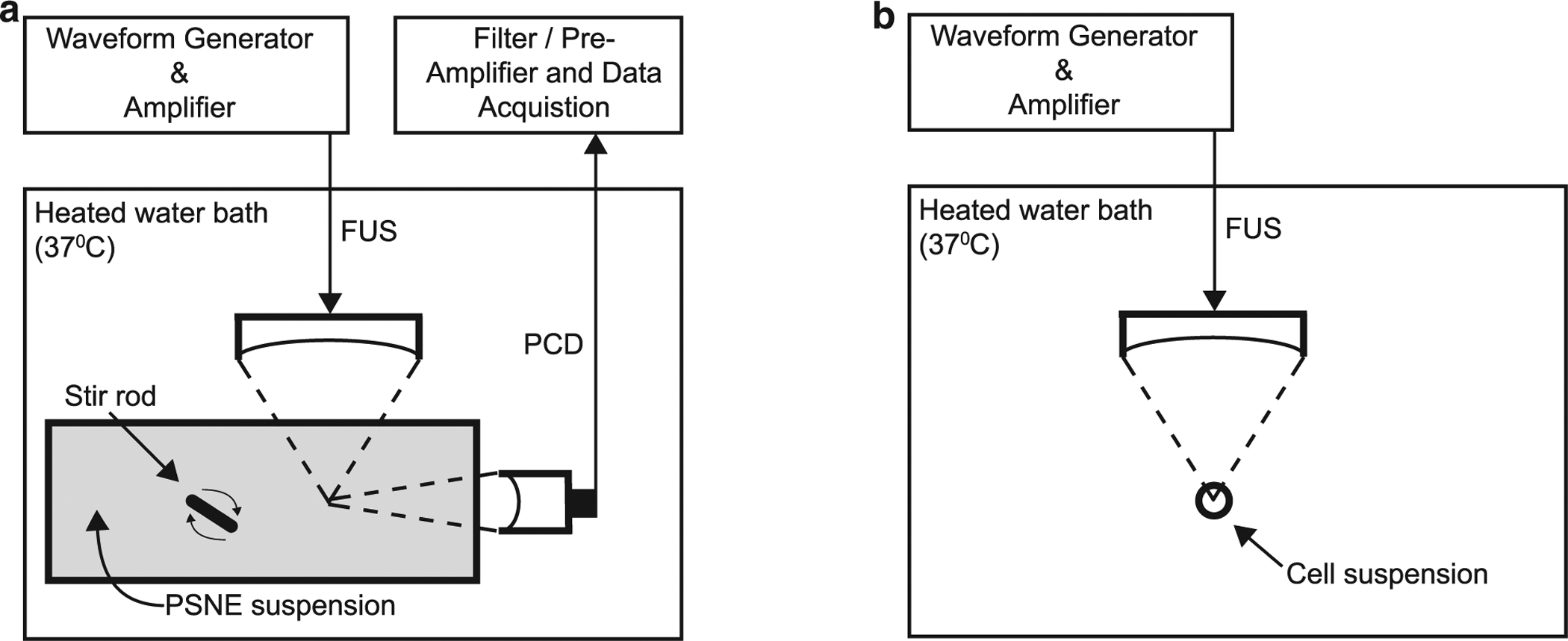
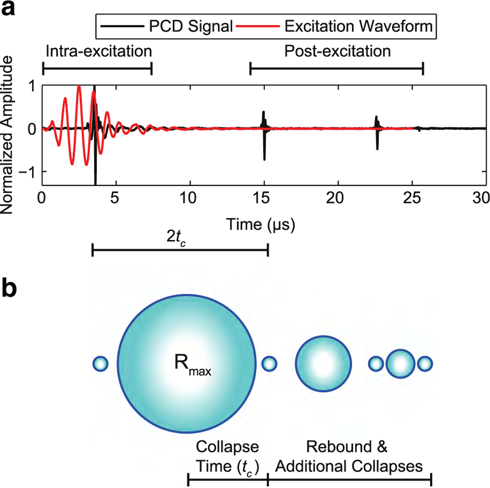
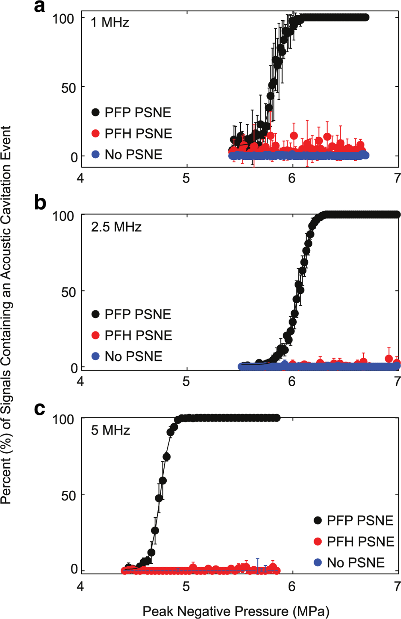
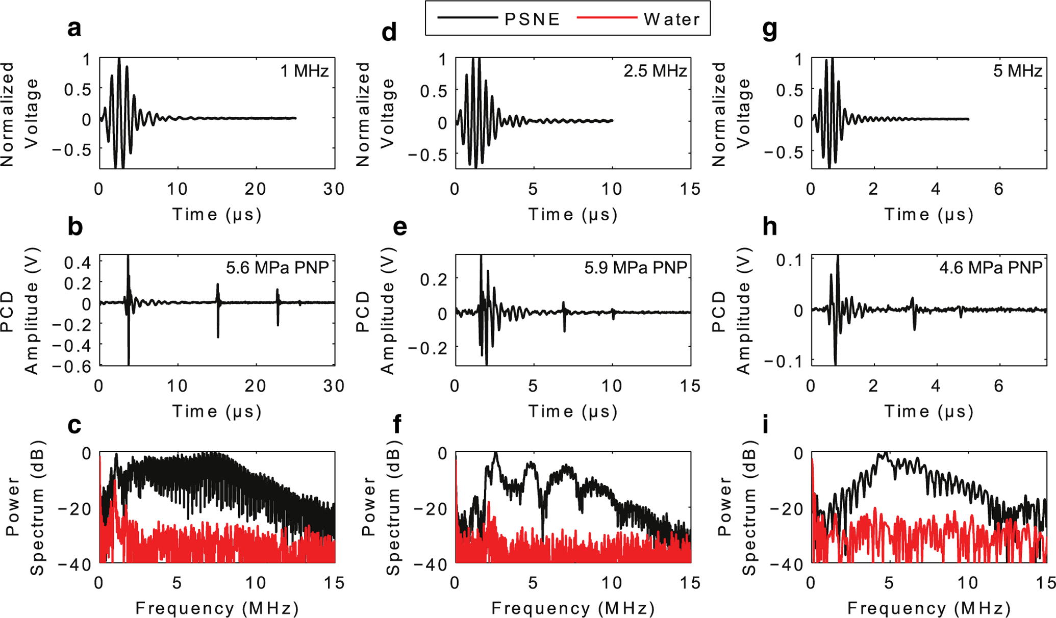
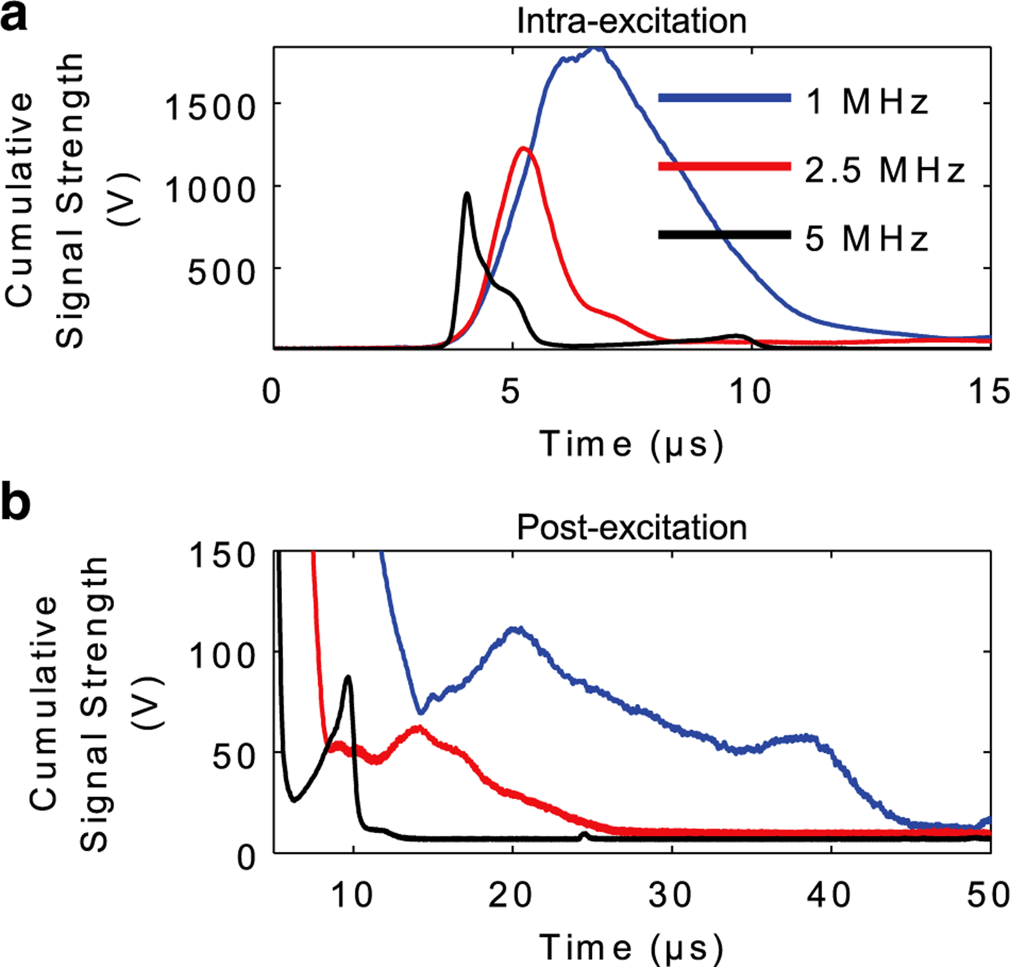
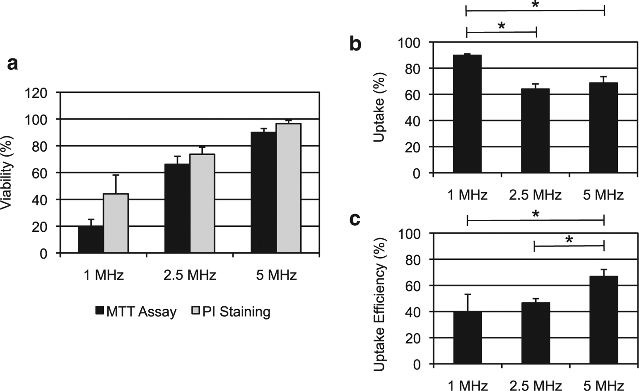
Acoustic cavitation can be used to temporarily disrupt cell membranes for intracellular delivery of large biomolecules. Termed sonoporation, the ability of this technique for efficient intracellular delivery (i.e., >50% of initial cell population showing uptake) while maintaining cell viability (i.e., >50% of initial cell population viable) has proven to be very difficult. Here, we report that phase-shift nanoemulsions (PSNEs) function as inertial cavitation nuclei for improvement of sonoporation efficiency. The interplay between ultrasound frequency, resultant microbubble dynamics and sonoporation efficiency was investigated experimentally. Acoustic emissions from individual microbubbles nucleated from PSNEs were captured using a broadband passive cavitation detector during and after acoustic droplet vaporization with short pulses of ultrasound at 1, 2.5 and 5 MHz. Time domain features of the passive cavitation detector signals were analyzed to estimate the maximum size (Rmax) of the microbubbles using the Rayleigh collapse model. These results were then applied to sonoporation experiments to test if uptake efficiency is dependent on maximum microbubble size before inertial collapse. Results indicated that at the acoustic droplet vaporization threshold, Rmax was approximately 61.7 ± 5.2, 24.9 ± 2.8, and 12.4 ± 2.1 μm at 1, 2.5 and 5 MHz, respectively. Sonoporation efficiency increased at higher frequencies, with efficiencies of 39.5 ± 13.7%, 46.6 ± 3.28% and 66.8 ± 5.5% at 1, 2.5 and 5 MHz, respectively. Excessive cellular damage was seen at lower frequencies because of the erosive effects of highly energetic inertial cavitation. These results highlight the importance of acoustic cavitation control in determining the outcome of sonoporation experiments. In addition, PSNEs may serve as tailorable inertial cavitation nuclei for other therapeutic ultrasound applications.

Keywords: Acoustic cavitation; Drug delivery; Inertial cavitation; Microbubbles; Sonoporation; Ultrasound.


Figures

Fig. 1.
Fig. 1.
Experimental setups used for detecting frequency-dependent acoustic emissions (a) during and after ADV of PSNEs and (b) frequency-dependent sonoporation efficiency. (a) FUS transducers were submerged in a heated bath of degassed, de-ionized water and used to interrogate a PSNE suspension (109 PSNEs/mL) with short pulses of FUS (5 cycles, 4–8 MPa peak negative pressure and 100-Hz pulse repetition frequency). Acoustic cavitation emissions were captured with a broadband PCD and post-processed to understand the dynamics of microbubbles derived from PSNEs after ADV. (b) A cell suspension containing cancer cells, PSNEs (109/mL) and mock biomolecule FITC-dextran (10 μM) were placed in a microcentrifuge tube and submerged in a heated bath of degassed, de-ionized water. FUS (5 cycles, 6.5-MPa peak negative pressure, 250-Hz pulse repetition frequency and 100-s treatment duration) was then used to vaporize PSNEs and promote acoustic cavitation activity within the cell suspension for intracellular delivery of FITC-dextran. ADV = acoustic droplet vaporization; FITC = fluorescein isothiocyanate; FUS = focused ultrasound; PCD = passive cavitation detector; PSNE = phase-shift nanoemulsions.
Fig. 2.
Fig. 2.
(a) An example time trace from the passive cavitation detector revealing intra- and post-excitation acoustic emissions relative to the focused ultrasound excitation waveform during the acoustic droplet vaporization of a single phase-shift nanoemulsion. The excitation waveform was a 1-MHz pulse at a peak negative pressure of 5.6 MPa. The waveform was measured using a calibrated hydrophone at a lower peak negative pressure (~1 MPa). (b) Hypothesized microbubble dynamics in relation to the temporal characteristics of the time trace. It is theorized that the microbubbles undergo rapid growth to a maximum radius (Rmax), followed by multiple collapses and rebounds after acoustic droplet vaporization of phase-shift nanoemulsions. The collapse time (tc) is defined as the time between the first intra-excitation collapse spike to the first post-excitation collapse spike.
Fig. 3.
Fig. 3.
(a-c) Acoustic droplet vaporization (ADV) threshold of phase-shift nanoemulsions (PSNE) as a function of the focused ultrasound (FUS) peak negative pressure at 1 MHz (a), 2.5 MHz (b), and 5 MHz (c). Using the experimental set-up in Figure 1a, a PSNE suspension (109 PSNE/mL) was interrogated with pulsed FUS (5 cycles, 4–8 MPa peak negative pressure and 100-Hz pulse repetition frequency), and acoustic cavitation emissions were simultaneously recorded with a broadband passive cavitation detector. PFP and PFH PSNEs were interrogated along with water (no PSNE) as a control. Results are depicted as the percentage of signals containing an acoustic cavitation event versus the peak negative pressure of the focused ultrasound. A total of 100 individual time traces were captured at each pressure step for post-processing. The ADV threshold was defined as the peak negative pressure that resulted in 10% of the signals containing acoustic cavitation events and was 5.6, 5.9 and 4.6 MPa at 1, 2.5 and 5 MHz, respectively. PFH = perfluorohexane; PFP = perfluropentane; PSNEs = phase-shift nanoemulsions.
Fig. 4.
Fig. 4.
Acoustic emissions from individual PSNEs after acoustic droplet vaporization with focused ultrasound at frequencies of (a–c) 1 MHz, (d–f) 2.5 MHz and (g–i) 5 MHz. (a, d, g) Focused ultrasound waveform for each frequency as measured with a needle hydrophone at a peak negative pressure of ~1 MPa. (b, e, h) Recorded time traces detected by the passive cavitation detector during and after acoustic droplet vaporization of single PSNEs. (c, f, i) Frequency content of the recorded time traces. The focused ultrasound pulse parameters were 5 cycles at peak negative pressures of 5.6, 5.9 and 4.6 MPa for 1, 2.5 and 5 MHz, respectively. PNP = peak negative pressure; PSNEs = phase-shift nanoemulsions.
Fig. 5.
Fig. 5.
Cumulative acoustic cavitation emissions during mock sonoporation experiments. PSNEs (109/mL) were interrogated with focused ultrasound using the experimental setup in Figure 1a and FUS parameters used in sonoporation experiments (5-cycle pulse, 6.5-MPa peak negative pressure, 250-Hz pulse repetition frequency and 100-s treatment duration). Cumulative signal strength from intra-excitation and post-excitation emissions was isolated by summing up the amplitude envelope of all 25,000 individual time traces. This provides information related to when acoustic cavitation activity occurred in time (i.e., collapse times) and the strength of that activity (i.e., cumulative amplitude). PSNE = phase-shift nanoemulsion.
Fig. 6.
Fig. 6.
Relationship between the FUS frequency and outcome of sonoporation experiments using PSNEs. The FUS parameters were fixed at 5 cycles, 6.5-MPa peak negative pressure, 250-Hz pulse repetition frequency and 100-s exposure duration. (A) Cell viability measured using a cell proliferation assay 24 h after focused ultrasound exposure (MTT assay) or immediately after the exposure with flow cytometry and PI staining. (B) Percentage of viable cells exhibiting uptake of mock biomolecule fluorescein isothiocyanate-dextran (20 kDa) after FUS exposure. (C) Uptake efficiency, which is the percentage of viable cells with uptake relative to the total number of initial cells, at the three FUS frequencies. FUS = focused ultrasound; PI = propidium iodide; PSNEs = phase-shift nanoemulsions.

References

    1. Ammi AY, Cleveland RO, Mamou J, Wang GI, Bridal SL, O’Brien WD. Ultrasonic contrast agent shell rupture detected by inertial cavitation and rebound signals. IEEE Trans Ultrason Ferroelectr Freq Control 2006;53:126–136. - PMC - PubMed
    1. Apfel RE. A novel technique for measuring the strength of liquids. J Acoust Soc Am 1971;49:145.
    1. Apfel R Acoustic cavitation prediction. J Acoust Soc Am 1981;69: 1624–1633.
    1. Apfel RE, Holland CK. Gauging the liklihood of cavitation from short-pulse, low-duty cycle diagnostic ultrasound. Ultrasound Med Biol 1991;17:179–185. - PubMed
    1. Avedisian CT. The homogeneous nucleation limits of liquids. J Phys Chem Ref Data 1985;14:695–729.

Publication types