. 2019 Jan 24;176(3):448-458.e12.
doi: 10.1016/j.cell.2018.11.040. Epub 2019 Jan 10.

Structure of a Signaling Cannabinoid Receptor 1-G Protein Complex

Affiliations

Structure of a Signaling Cannabinoid Receptor 1-G Protein Complex

Kaavya Krishna Kumar et al. Cell. .

Abstract

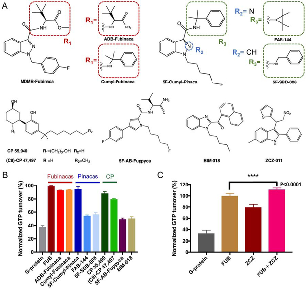
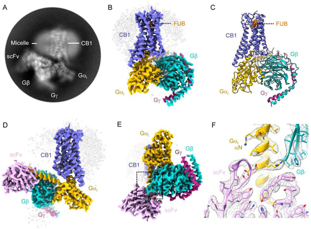
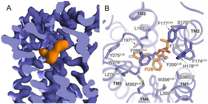
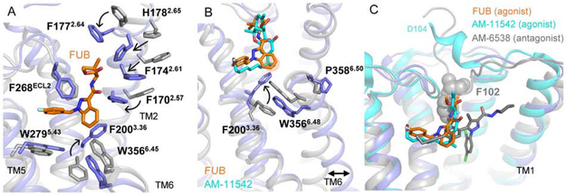
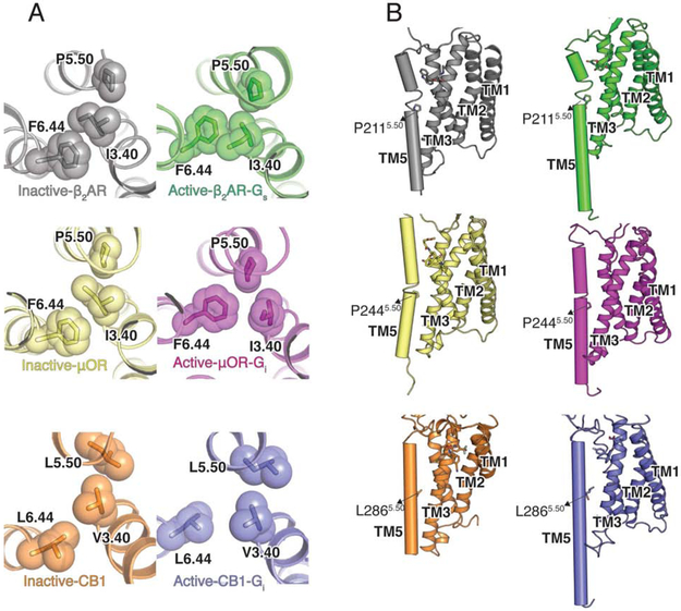
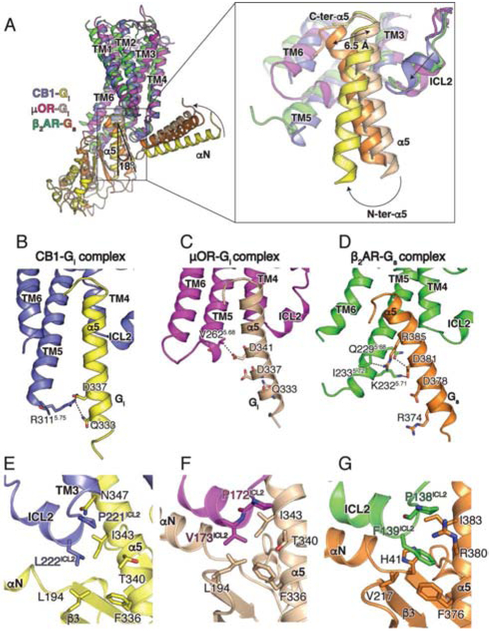
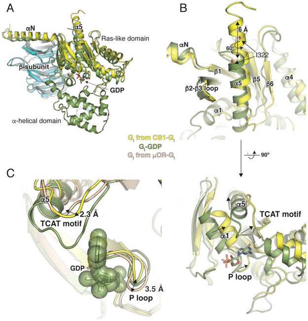
Cannabis elicits its mood-enhancing and analgesic effects through the cannabinoid receptor 1 (CB1), a G protein-coupled receptor (GPCR) that signals primarily through the adenylyl cyclase-inhibiting heterotrimeric G protein Gi. Activation of CB1-Gi signaling pathways holds potential for treating a number of neurological disorders and is thus crucial to understand the mechanism of Gi activation by CB1. Here, we present the structure of the CB1-Gi signaling complex bound to the highly potent agonist MDMB-Fubinaca (FUB), a recently emerged illicit synthetic cannabinoid infused in street drugs that have been associated with numerous overdoses and fatalities. The structure illustrates how FUB stabilizes the receptor in an active state to facilitate nucleotide exchange in Gi. The results compose the structural framework to explain CB1 activation by different classes of ligands and provide insights into the G protein coupling and selectivity mechanisms adopted by the receptor.

Keywords: CB1; Fubinaca; G(i); GPCR; cannabinoid receptor; cryo-EM; synthetic cannabinoid.


Figures

Figure 1.
Figure 1.. GTP turnover assay for the CB1 ligands.
(A) Chemical structures of CB1 agonists used in this study, and of PAM ZCZ. (B) GTP turnover assay for the 10 ligands tested. (C) The addition of ZCZ further increases GTP turnover indicating PAM activity. Data are normalized to FUB in (B) and (C).
Figure 2.
Figure 2.. Cryo-EM structure of the CB1-Gi complex.
(A) Representative reference-free two-dimensional (2D) cryo-EM average of CB1-FUB-Gi-scFv16 shows high resolution features including the helical pitch of TM α-helices. The labels indicate complex components. The diameter of the circular mask is 18 nm. (B-E) Three-dimensional map (B, D-E) and model (C) obtained from cryo-EM of the CB1-Gi complex. CB1 is colored blue, FUB - orange, Gαi, −β and −γ yellow, cyan and dark magenta, respectively, and scFv16 is colored pink. (F) Snapshot of model vs. map density in the region where the ScFv16 is engaging Gαi and Gβ. The zoomed in region corresponds to the area highlighted by a dashed black box in (E).
Figure 3.
Figure 3.. The FUB binding pocket.
(A) Cut-through view of CB1 cryo-EM map with FUB bound in the orthosteric pocket. Density corresponding to FUB is colored orange, and CB1 purple. (B) FUB interactions in the CB1 binding site.
Figure 4.
Figure 4.. CB1 activation by FUB.
(A) Superposition of the FUB activated complex (blue) with an inverse agonist bound receptor (AM-6358, PDB 5TGZ, grey). (B) FUB and AM-11542 (PDB 5XRA) bound at the CB1 orthosteric pocket make direct contacts with residues F2003.36 and W3566.48. The rotation of F2003.36 to interact with the indazole ring of FUB allows W3566.48 to rotate outwards, with a consequent outward movement of the cytoplasmic end of TM6 that serves to create a cavity for G protein binding. The groups interacting with the ‘toggle twin switch’ of CB1 (indazole ring of FUB and GDH moiety of AM-11542) are marked in orange. The inactive receptor structure is shown in grey (PDB 5TGZ). (C) A comparison of binding pockets of AM-6538 (antagonist, grey, PDB 5TGZ), AM-11542 (agonist, cyan, PDB 5XRA) and FUB (orange). The receptor is presented as cartoon with the active conformation in blue (present cryo-EM structure) and the inactive conformation in gray (PDB 5TGZ).
Figure 5.
Figure 5.. Structural changes in CB1 on nucleotide-free Gi binding.
(A) Structural rearrangement of P-I-F motif in CB1, μOR (inactive- PDB 4DKL, yellow; PDB 6DDE, magenta) and β2AR (inactive-PDB 2RH1, grey; active-PDB 3SN6, green) upon activation. (B) The local unwinding of TM5 due to P5.50 in active and inactive β2AR (green) and μOR (magenta) is not seen in CB1.
Figure 6.
Figure 6.. Relative orientation of CB1 and Gi and Role of ICL2 in coupling selectivity.
(A) Comparison of the relative orientation of Gi bound to CB1 (blue), μOR (PDB 6DDE, magenta), and β2AR (PDB 3SN6, green) when aligned on the receptor. A magnified view is provided of the position of the α5 helices in CB1-Gαi (yellow), μOR-Gαi (wheat) and β2AR-Gαs (orange). (B, C, D) Residues in the TM5-TM6 helices of CB1 (B), μOR (C) and β2AR (D) interacting with the α5 helix of Gαi (bound to CB1, yellow and bound to μOR, wheat) and Gαs (orange). (E, F, G) Interactions between ICL2 of CB1 (blue) and Gαi (yellow), μOR (magenta) and Gαi (wheat) and β2AR (green) and Gαs (orange).
Figure 7.
Figure 7.. Structural changes in Gi on CB1 binding.
(A) Comparison of GDP-bound Gαi (PDB 1GP2, green) and nucleotide-free Gαi from the CB1-Gi complex (yellow). The structures are aligned on the β-subunit (CB1-Gi, cyan and GDP-bound Gαi, grey). GDP is shown as sticks. The alpha helical domain (AHD) seen in the GDP-bound structure is not resolved in the nucleotide-free Gαi bound to CB1. (B) The α5 helix of Gαi moves upward by 6 Å and rotates ~ 60° to engage the receptor core. The TCAT motif that coordinates the guanosine base of GDP in the GDP-bound structure (green) has shifted upwards in the nucleotide-free Gαi bound to μOR (wheat). In CB1-bound Gαi (yellow), the TCAT motif is in a similar position as that seen in A2A-bound mini-Gαs (with GDP) (PDB 5G53, purple). (C) The conformation of the P loop in CB1-bound Gi (yellow) will allow nucleotide binding as seen when overlayed with GDP-bound Gi (1GP2, smudge). However in the nucleotide-free G protein bound to μOR (μOR-Gi, sand), there is a clash of the P loop with the GDP.

Comment in

References

    1. Adams AJ, Banister SD, Irizarry L, Trecki J, Schwartz M, and Gerona R (2017). “Zombie” Outbreak Caused by the Synthetic Cannabinoid AMB-FUBINACA in New York. N. Engl. J. Med 376, 235–242. - PubMed
    1. Adams PD, Afonine PV, Bunkóczi G, Chen VB, Echols N, Headd JJ, Hung L-W, Jain S, Kapral GJ, Grosse-Kunstleve RW, et al. (2011). The Phenix software for automated determination of macromolecular structures. Methods 55, 94–106. - PMC - PubMed
    1. Afonine PV, Klaholz BP, Moriarty NW, Poon BK, Sobolev OV, Terwilliger TC, Adams PD, and Urzhumtsev A (2018). New tools for the analysis and validation of Cryo-EM maps and atomic models. 1–66. - PMC - PubMed
    1. Atwood BK, Huffman J, Straiker A, and Mackie K (2010). JWH018, a common constituent of “Spice” herbal blends, is a potent and efficacious cannabinoid CB receptor agonist. Br. J. Pharmacol 160, 585–593. - PMC - PubMed
    1. Ballesteros JA, and Weinstein H (1995). Integrated methods for the construction of three-dimensional models and computational probing of structure-function relations in G protein-coupled receptors. In Methods in Neurosciences, (Elsevier), pp. 366–428.

Publication types

MeSH terms