

Multimodal dopaminergic and free-water imaging in Parkinson's disease
- PMID: 30639168
- PMCID: PMC6589363
- DOI: 10.1016/j.parkreldis.2019.01.007
Multimodal dopaminergic and free-water imaging in Parkinson's disease
Abstract
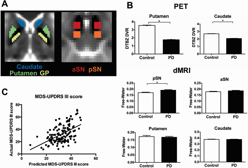
Introduction: When using free-water diffusion imaging or positron emission tomography (PET), it is established that substania nigra microstructure and presynaptic dopamine activity are impaired in early PD. It is not well understood if these two forms of degeneration are redundant, or if they each provide a unique contribution to the clinical motor and cognitive symptoms.
Methods: A total of 129 PD and 75 control individuals underwent motor and cognitive evaluations, and in vivo [11C]dihydrotetrabenazine (DTBZ) monoaminergic brain PET imaging and diffusion imaging. Correlations between free-water in the substantia nigra and striatal PET measures were analyzed. Unbiased multiple regression using a backward elimination method was performed between clinical severity and all imaging measures.
Results: Inverse correlations were found between free-water in posterior substantia nigra and DTBZ binding in putamen and caudate. Multiple regression revealed that increased free-water in the posterior substantia nigra, decreased DTBZ binding in putamen, and age were predictors of higher Hoehn and Yahr stage, MDS-UPDRS III scores, and posture and gait sub-scores. Increased posterior substantia nigra free-water alone was associated tremor scores. Free-water in caudate and putamen did not predict measures of motor performance. Decreased DTBZ binding in caudate, increased free-water in caudate and posterior substantia nigra were associated with higher dementia ratings.
Conclusions: These findings suggest that free-water in the posterior substantia nigra and presynaptic dopamine imaging in striatum are uniquely associated with the clinical symptoms of PD, indicating that each imaging modality may be measuring a unique mechanism relevant to nigrostriatal degeneration.
Keywords: Basal ganglia; Diffusion magnetic resonance imaging; Multimodal imaging; PET; Parkinson's disease; Substantia nigra.
Copyright © 2019 Elsevier Ltd. All rights reserved.
Figures
References
-
- Pringsheim T, Jette N, Frolkis A, Steeves TD. The prevalence of Parkinson’s disease: a systematic review and meta-analysis. Movement disorders : official journal of the Movement Disorder Society. 2014;29:1583–90. - PubMed
-
- Kalia LV, Lang AE. Parkinson’s disease. Lancet (London, England). 2015;386:896–912. - PubMed
-
- Pasternak O, Sochen N, Gur Y, Intrator N, Assaf Y. Free water elimination and mapping from diffusion MRI. Magnetic resonance in medicine. 2009;62:717–30. - PubMed
Publication types
MeSH terms
Substances
Grants and funding
LinkOut - more resources
Full Text Sources
Medical
Research Materials
Miscellaneous
