Review
. 2019 Mar 5;27(3):420-426.
doi: 10.1016/j.str.2018.11.011. Epub 2019 Jan 10.

HIV-1 Reverse Transcriptase: A Metamorphic Protein with Three Stable States

Affiliations
Review

HIV-1 Reverse Transcriptase: A Metamorphic Protein with Three Stable States

Robert E London. Structure. .

Abstract

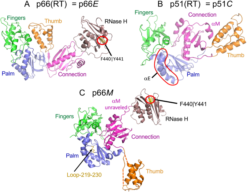
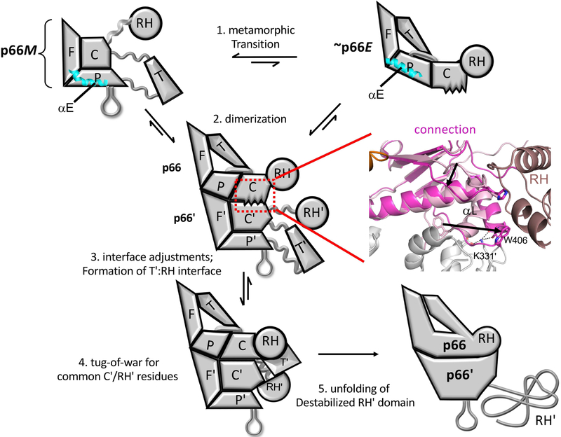
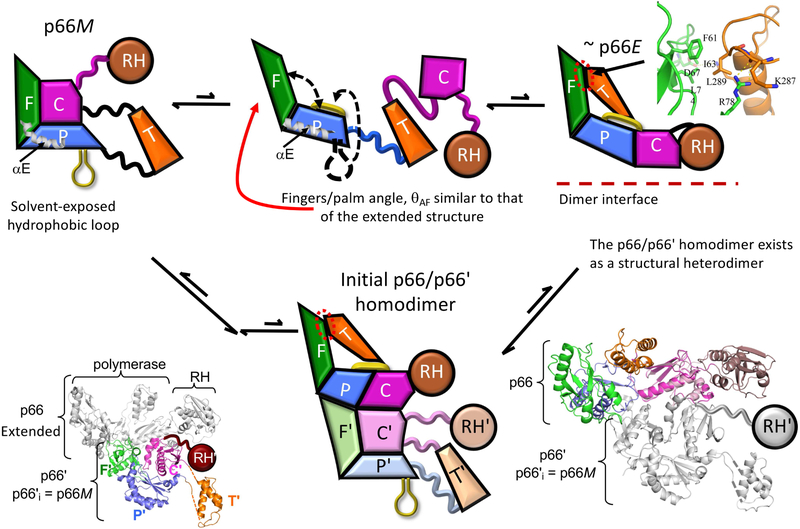
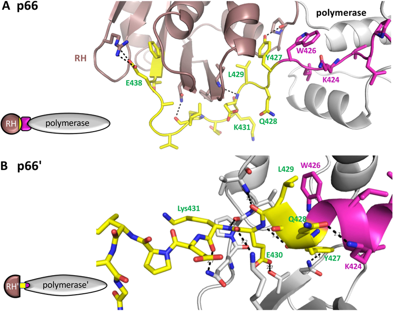
There has been a steadily increasing appreciation of the fact that the relationship between protein sequence and structure is often sufficiently ambiguous to allow a single sequence to adopt alternative, stable folds. Living organisms have been able to utilize such metamorphic proteins in remarkable and unanticipated ways. HIV-1 reverse transcriptase is among the earliest such proteins identified and remains a unique example in which a functional heterodimer contains two, alternatively folded polymerase domains. Structural characterization of the p66 precursor protein combined with NMR spectroscopic and molecular modeling studies have provided insights into the factors underlying the metamorphic transition and the subunit-specific programmed unfolding step required to expose the protease cleavage site within the ribonuclease H domain, supporting the conversion of the p66/p66' precursor into the mature p66/p51 heterodimer.

Keywords: HIV-1 reverse transcriptase; metamorphic protein; metamorphic transition; programmed unfolding; subunit-specific RH-domain unfolding.


Figures

Figure 1.
Figure 1.. Alternate structures of the RT polymerase domain.
Ribbon diagrams of the HIV-1 RT p66 subunit (A), the p51 subunit (B), and the p66 monomer (p66M) (C). The polymerase subdomains are color coded: fingers (green), palm (blue), thumb (orange), connection (magenta), and the RH domain is brown. The polymerase domain in the p66 subunit contains a short β-sheet formed from the palm domain C-terminus and the connection domain N-terminus that is absent in the monomer and the p51 subunit. The structure of the polymerase domain in p66M is fairly similar to that of p51C, with the major differences being the unfolding of helix αM and the disorder of segments connecting the thumb to the palm and connection subdomains, so that its orientation is variable. The F440∣Y441 cleavage site (yellow) in the RH domain in both the extended structure (A) and the p66 monomer (C) is inaccessible to the solvent and to the protease.
Figure 2.
Figure 2.. Schematic illustration of the main steps involved in RT maturation.
Step 1: Dissociation of the connection domain from the fingers/palm is followed by a series of conformational changes that constitute the metamorphic transition of the polymerase domain, altering the structure to a family of p66E-like forms, that can dimerize with the predominant p66M species. 2. an initial dimer is formed, followed by conformational adjustments at the interface that involve primarily the connection and palm subdomains. The ribbon diagram to the right illustrates some of the necessary conformational adjustments of the connection subdomain. 3. The subunit interface between the thumb' and the RH domain is formed, and the unfolded residues from the C-terminus of the p66' connection' domain occupy the channel between the connection' and thumb' domains. 4. Inherent instability of RH' domain releases Tyr427' and adjacent residues that are then captured by an extending helix αM'. 5. The destabilized RH' domain unfolds, exposing the cleavage site. Straightening of α-helix E in the palm subdomain (cyan) is one of the drivers for the metamorphic transition.
Figure 3.
Figure 3.. Drivers for the metamorphic transition.
After dissociation of the connection subdomain, the fingers/palm subdomains rotate away from each other, and the palm C-terminus forms a short, amphiphilic β-sheet that lies against the palm domain β-sheet (β6-β9-β10). The tips of the fingers and thumb form a weak interface that helps to position the system for dimer formation. A ribbon diagram of the fingers – thumb interface is shown at the upper right. The initial p66/p66' homodimer is a structural heterodimer that is formed from a p66E-like structure and the p66M monomer structure. The ribbon diagram to the lower left corresponds to the initial homodimer with color-coded p66' subdomains; in the ribbon diagram to the lower right, the p66 subdomains are color coded, and the overlaid initial connection domain conformation is indicated in light pink.
Figure 4.
Figure 4.. Subunit-specific "tug-of-war" for N-terminal RH domain residues.
In the p66 subunit (A), the C-terminal segment of the connection domain (magenta) adopts an extended conformation that links the polymerase (gray) and RH (brown) domains. The N-terminal segment of the RH domain from Tyr427-Tyr440 is colored yellow. In the p66' subunit (B), The polymerase domain C-terminal residues form a helix (magenta) that extends to include several residues derived from the RH' domain (yellow) when this domain is transiently unfolded. In the structure shown (pdb: 1RTJ, (Esnouf et al., 1995)), 8 hydrogen bonds are shown between residues derived from the RH' domain and the polymerase' domain, and additional interactions include the hydrophobic interface between Leu429 and Trp426. In both panels, the polymerase domain is gray, except for the C-terminal residues (magenta); the N-terminal RH domain residues Tyir427-Phe440 are yellow, and the remaining RH domain residues are brown. In panel B, the sidechain conformations of Q242 and Q428 were adjusted relative to structure 1RTJ to optimize the H-bond interactions. The schematics on the left illustrate the inclusion of RH' domain N-terminal residues (yellow) into the polymerase' domain.

References

    1. Abram ME, and Parniak MA (2005). Virion instability of human immunodeficiency virus type 1 reverse transcriptase (RT) mutated in the protease cleavage site between RT p51 and the RT RNase H domain. J Virol 79, 11952–11961. - PMC - PubMed
    1. Becerra SP, Kumar A, Lewis MS, et al. (1991). Protein-protein interactions of HIV-1 reverse transcriptase: implication of central and C-terminal regions in subunit binding. Biochemistry-Us 30, 11707–11719. - PubMed
    1. Belshaw R, Pybus OG, and Rambaut A (2007). The evolution of genome compression and genomic novelty in RNA viruses. Genome research 17, 1496–1504. - PMC - PubMed
    1. Braz VA, Holladay LA, and Barkley MD (2010). Efavirenz binding to HIV-1 reverse transcriptase monomers and dimers. Biochemistry-Us 49, 601–610. - PMC - PubMed
    1. Choi H, Kim S, Mukhopadhyay P, et al. (2001). Structural basis of the redox switch in the OxyR transcription factor. Cell 105, 103–113. - PubMed

Publication types

MeSH terms

Substances

LinkOut - more resources