Review
. 2019 Jan 1:138:302-325.
doi: 10.1016/j.addr.2019.01.005. Epub 2019 Jan 11.

Iron oxide nanoparticles: Diagnostic, therapeutic and theranostic applications

Affiliations
Review

Iron oxide nanoparticles: Diagnostic, therapeutic and theranostic applications

Seyed Mohammadali Dadfar et al. Adv Drug Deliv Rev. .

Abstract

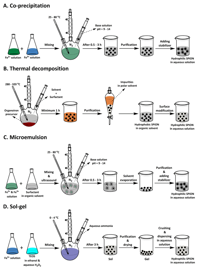
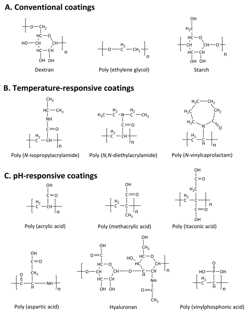
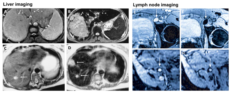
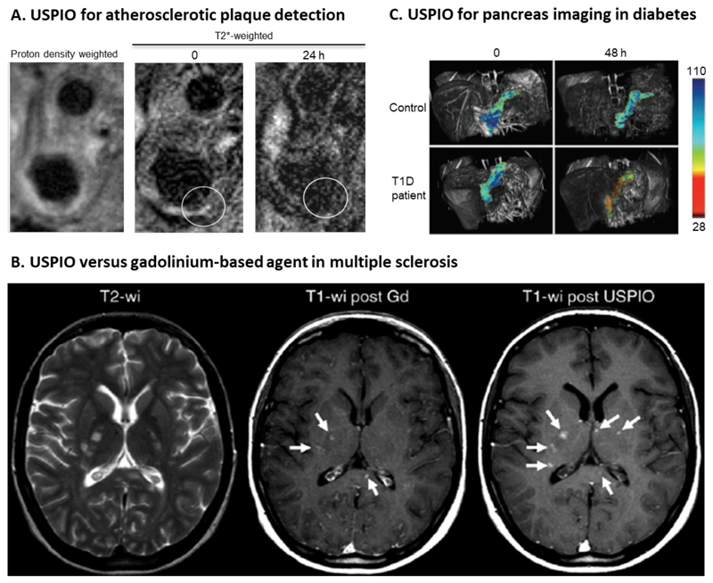
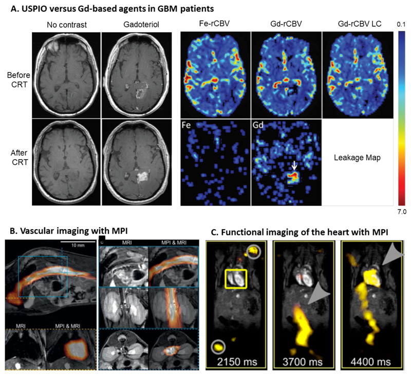
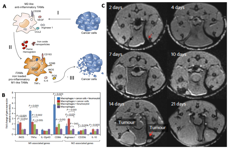
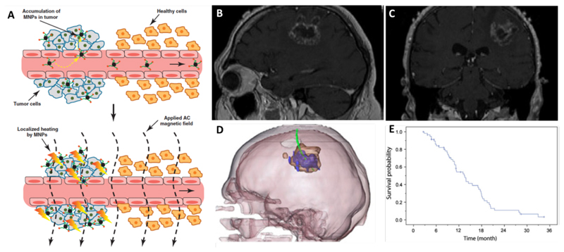
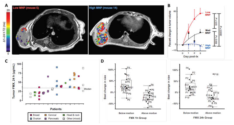
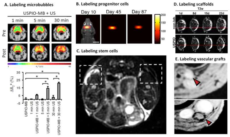
Many different iron oxide nanoparticles have been evaluated over the years, for a wide variety of biomedical applications. We here summarize the synthesis, surface functionalization and characterization of iron oxide nanoparticles, as well as their (pre-) clinical use in diagnostic, therapeutic and theranostic settings. Diagnostic applications include liver, lymph node, inflammation and vascular imaging, employing mostly magnetic resonance imaging but recently also magnetic particle imaging. Therapeutic applications encompass iron supplementation in anemia and advanced cancer treatments, such as modulation of macrophage polarization, magnetic fluid hyperthermia and magnetic drug targeting. Because of their properties, iron oxide nanoparticles are particularly useful for theranostic purposes. Examples of such setups, in which diagnosis and therapy are intimately combined and in which iron oxide nanoparticles are used, are image-guided drug delivery, image-guided and microbubble-mediated opening of the blood-brain barrier, and theranostic tissue engineering. Together, these directions highlight the versatility and the broad applicability of iron oxide nanoparticles, and indicate the integration in future medical practice of multiple iron oxide nanoparticle-based materials.

Keywords: MFH; MPI; MRI; Nanomedicine; SPION; Theranostics; USPIO.


Figures

Figure 1
Figure 1. Schematic depiction of commonly used chemical strategies to prepare iron oxide nanoparticles.
A: Co-precipitation, B: Thermal decomposition, C: Micro-emulsion and D: Sol-gel. In terms of simplicity, the co-precipitation technique is the most suitable. In terms of size and morphology control, and for producing SPION smaller than 20 nm, thermal decomposition and microemulsion are preferred. The sol-gel technique is useful to produce SPION with silica coatings and with a size larger than 20 nm.
Figure 2
Figure 2. Examples of polymers used as surface coatings of iron oxide nanoparticles.
Conventional polymers (A) are used as coatings in the commercial iron oxide nanoparticle formulations listed in Table 1. For certain applications, iron oxide nanoparticles are surface-functionalized with temperature (B) or pH (C) -responsive polymeric coating.
Figure 3
Figure 3. SPION for liver imaging and lymph node imaging.
A-B: T2-weighted MR image of a liver with a large hepatocellular carcinoma before (A) and after (B) the administration of SPION. The lesion is demarcated with arrows [168]. C-D: Standard (C) and SPION-based contrast-enhanced (D) MR imaging of liver metastasis in a patient with colorectal cancer. After administration of ferumoxide SPION, a second metastasis becomes visible on T2-weighted MR image [148]. E-H: Lymph node in left iliac region (arrow), with and without metastatic infiltration. T2-weighted images before (E, G) and 24 h after (F, H) administration of ferumoxtran. Lymph node (arrow) appears bright before injection of UPIO (E, G). One day after injection, a signal loss in the lymph node (arrow) due to high UPIO macrophage uptake can be observed, thus indicating functionality and no metastasis (F). Conversely, in the lower panel, the lymph node (arrow) stays bright, indicating no trafficking of USPIO and thus metastatic colonization (H) [161].
Figure 4
Figure 4. Imaging inflammation with USPIO nanoparticles.
A: MR images of external and internal carotid artery in a patient with atherosclerosis. T2*-weighted MR images before (middle) and 24 h after (right) the intravenous administration of USPIO nanoparticles. Around the vessel wall (circle) a decrease in signal intensity can be observed [172] B: Differences in enhancement pattern between USPIO nanoparticles and gadolinium-based MRI contrast agents in a patient suffering from multiple sclerosis (MS). Left, multiple hyperintense lesions can be detected on the non-contrast-enhanced T2-weighted image. Middle, gadolinium-based T1-weighted image showing three contrast-enhanced MS lesions (arrows). USPIO with core diameter smaller than 4 nm show shortening of T1 relaxation and therefore have been used for T1-weighted images. Right, T1-weighted image obtained after USPIO administration, showing the same three lesions and three additional lesions (arrows), exemplifying the added value of iron oxide-based diagnostic assessment in MS [175]. C: T2-based pseudocolor MR images showing enhanced SPION accumulation in a T1D diabetic patient as compared to a healthy individual [178].
Figure 5
Figure 5. Iron oxide nanoparticles for vascular imaging.
[184]. A: Non-contrast-enhanced and gadolinium-enhanced Tl-weighted MR images before and after chemoradiotherapy (CRT) show an increase in hyperintensity in the lesion, indicative of (pseudo-) progression. Relative cerebral blood volume (rCBV) evaluated with ferumoxytol (Fe-rCBV), gadoteridol (Gd-rCBV) and gadoteridol with leakage correction (Gd-rCBV LC) all show low rCBV, which identifies this situation as pseudoprogression. The leakage map shows contrast agent accumulation for gadoteridol (arrow), while no leakage is observed for ferumoxytol [191]. B: Images combining magnetic particle imaging (MPI) with MRI. Circulating ferucarbotran-based SPION are visualized by MPI, while MRI provides anatomical information. Bottom left images show SPION circulating through the heart. The top images and bottom right images show SPION in the inferior vena cava (sagittal, coronal and transverse orientation, respectively) [194]. C: In vivo measurement from a beating mouse heart by MRI and overlaid with traveling wave MPI data. At 2150 m after the i.v. injection ferucarbotran-based SPION, no signal can be detected in the heart (yellow box), only the signal of the marker points for co-registration (grey circle) is detectable. At 3700 ms, the SPION can be seen in the artery that is leading to the heart (grey arrow), and after 4400 ms, the SPION eventually reaches the heart (grey arrow). [195].
Figure 6
Figure 6. Iron oxide nanoparticles alter macrophage polarization towards an anti-tumoral phenotype.
A. SPION or hemoglobin induce a shift in activation of tumor growth supporting TAM to a more M1-like anti-tumoral phenotype. I. Tumor cells support M2-like polarization by inducing a variety of growth factors and receptors like CCL2, VEGF or cluster of differentiation 206 (CD206). II. TAM exposed to SPION or hemoglobin change their polarization towards a pro-inflammatory phenotype with high generation of TNFα, IL-6 and ROS. III. Iron-loaded TAM show an anti-tumoral activity and decrease the viability of cancer cells [211]. B. Polarization of macrophages induced by ferumoxytol. PCR results show a significant increase in the expression of typical M1 markers like TNFα for the iron oxide nanoparticle-treated group compared to untreated. C. Co-injection of SPION (ferumoxtran or ferumoxytol) with MMTV-PyMT derived cancer cells into the mammary fat pad of mice showed a significant inhibition of tumor growth in comparison to the non-SPION containing controls. T2*-weighted MR images on day 2 shows SPION-related darkening (red arrow) which is absent on the side which was only injected with cancer cells. On day 14 after tumor cell inoculation, MRI shows a growth inhibitory effect for ferumoxytol co-injection (red arrow) as compared to controls (black arrow) [214].
Figure 7
Figure 7. Magnetic drug targeting and magnetic fluid hyperthermia.
A. Schematic depiction of magnetic fluid hyperthermia mechanism of action. The upper image shows the specific accumulation of magnetic nanoparticles (MNP) in tumor tissue due to leaky vasculature (EPR). The lower image exemplifies that after the application of an alternating current (AC) magnetic field, the SPION which have accumulated in the tumor heat up and lead to tissue damage [218]. B-C. MR images of patients with recurrent GBM before treatment. D. Fusion of MRI and CT (3D-reconstruction) shows the thermometry catheter in green, magnetic fluid in blue and glioblastoma tumor mass in brown. E. Overall survival of 59 glioblastoma patients after diagnosis of first tumor recurrence and treatment with combination of magnetic fluid hyperthermia and radiotherapy [219].
Figure 8
Figure 8. Iron oxide-based companion diagnostics.
A: T2-weigthed MR images (overlaid with pseudocolor images indicating ΔT2 in the tumor area) show varying levels of accumulation of ferumoxytol-based iron oxide nanoparticles in HT1080 tumors in mice. Low and high magnetic nanoparticle (MNP) corresponds to tumors with low and high levels of tumor targeting. B: Correlation between the tumor accumulation of iron oxide-based companion diagnostics and the antitumor response progression induced by paclitaxel-loaded PLGA-PEG nanoparticles [235]. C: Ferumoxytol concentration in different tumor lesions at 24 h after i.v. injection, showing highly heterogeneous uptake in individual lesions. D: Correlation between lesion size change after treatment with irinotecan-loaded liposomes determined by CT measurements on the one hand, and ferumoxytol uptake in the different lesions detected by MRI at 1 h and 24 h after injection on the other hand. For every time point, the median tumor accumulation of ferumoxytol was calculated. Based on this, the lesions were classified in below vs. above the median, and this was correlated with irinotecan-loaded liposome treatment efficacy. A decent correlation between ferumoxytol uptake and lesion size change was observed [236].
Figure 9
Figure 9. Theranostic applications of iron oxide nanoparticles in brain drug delivery and in regenerative medicine.
A: Permeation of the blood-brain barrier (BBB), induced and imaged using microbubbles of which the shell was loaded with iron oxide nanoparticles (USPIO-MB). Upper panel: T2*-weighted images overlaid with color-coded R*-maps, showing ultrasound-induced destruction of USPIO-MB, BBB opening and the subsequent deposition of iron oxide nanoparticles in the brain. Lower panel: Quantification of R2* values shows gradual enhancement of BBB opening when increasing the time of ultrasound exposure [247]. B: Tracing of ferucarbotran-labeled human neuronal progenitor cells with MPI in the forebrain cortex of rats [198]. C: SPION-labeled mesenchymal stem cells (MSC) in collagen-based hydrogels 19 days after implantation, visualized using MRI. T2*-weighted image shows hypointense region on right side compared to the unlabeled control implanted on left side. [256]. D: Unlabeled (upper) and USPIO-labeled (lower) collagen scaffolds implanted in mice, showing clear dark areas in T2-weighted MRI in case of iron oxide nanoparticle labeling [257]. E: Tissue-engineered vascular grafts implanted as a shunt between the carotid artery and the jugular vein in sheep can be clearly visualized using proton density-weighted MRI when iron oxide nanoparticles are integrated (compare lower vs. upper panel) [258].

References

    1. Figuerola A, Di Corato R, Manna L, Pellegrino T. From iron oxide nanoparticles towards advanced iron-based inorganic materials designed for biomedical applications. Pharmacol Res. 2010;62:126–143. - PubMed
    1. Mohammed L, Gomaa HG, Ragab D, Zhu J. Magnetic nanoparticles for environmental and biomedical applications: A review. Particuology. 2017;30:1–14.
    1. Wu W, Wu Z, Yu T, Jiang C, Kim W-S. Recent progress on magnetic iron oxide nanoparticles: synthesis, surface functional strategies and biomedical applications. Sci Technol Adv Mater. 2015;16 023501. - PMC - PubMed
    1. Jin R, Lin B, Li D, Ai H. Superparamagnetic iron oxide nanoparticles for MR imaging and therapy: design considerations and clinical applications. Curr Opin Pharmacol. 2014;18:18–27. - PubMed
    1. Cheng F-Y, Su C-H, Yang Y-S, Yeh C-S, Tsai C-Y, Wu C-L, Wu M-T, Shieh D-B. Characterization of aqueous dispersions of Fe3O4 nanoparticles and their biomedical applications. Biomaterials. 2005;26:729–738. - PubMed

Publication types