Randomized Controlled Trial
. 2019 Apr 1;76(4):420-429.
doi: 10.1001/jamaneurol.2018.4304.

Utility of Neuronal-Derived Exosomes to Examine Molecular Mechanisms That Affect Motor Function in Patients With Parkinson Disease: A Secondary Analysis of the Exenatide-PD Trial

Affiliations
Randomized Controlled Trial

Utility of Neuronal-Derived Exosomes to Examine Molecular Mechanisms That Affect Motor Function in Patients With Parkinson Disease: A Secondary Analysis of the Exenatide-PD Trial

Dilan Athauda et al. JAMA Neurol. .

Erratum in

  • Error in Byline.
    [No authors listed] [No authors listed] JAMA Neurol. 2019 Apr 1;76(4):509. doi: 10.1001/jamaneurol.2019.0575. JAMA Neurol. 2019. PMID: 30958554 Free PMC article. No abstract available.

Abstract

Importance: Exenatide, a glucagon-like peptide 1 agonist used in type 2 diabetes, was recently found to have beneficial effects on motor function in a randomized, placebo-controlled trial in Parkinson disease (PD). Accumulating evidence suggests that impaired brain insulin and protein kinase B (Akt) signaling play a role in PD pathogenesis; however, exploring the extent to which drugs engage with putative mechnisms in vivo remains a challenge.

Objective: To assess whether participants in the Exenatide-PD trial have augmented activity in brain insulin and Akt signaling pathways.

Design, setting, and participants: Serum samples were collected from 60 participants in the single-center Exenatide-PD trial (June 18, 2014, to June 16, 2016), which compared patients with moderate PD randomized to 2 mg of exenatide once weekly or placebo for 48 weeks followed by a 12-week washout period. Serum extracellular vesicles, including exosomes, were extracted, precipitated, and enriched for neuronal source by anti-L1 cell adhesion molecule antibody absorption, and proteins of interest were evaluated using electrochemiluminescence assays. Statistical analysis was performed from May 1, 2017, to August 31, 2017.

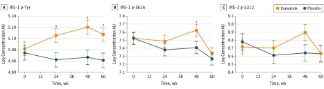
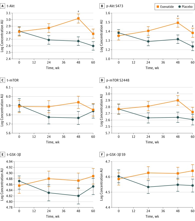
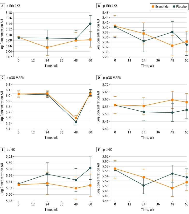
Main outcomes and measures: The main outcome was augmented brain insulin signaling that manifested as a change in tyrosine phosphorylated insulin receptor substrate 1 within neuronal extracellular vesicles at the end of 48 weeks of exenatide treatment. Additional outcome measures were changes in other insulin receptor substrate proteins and effects on protein expression in the Akt and mitogen-activated protein kinase pathways.

Results: Sixty patients (mean [SD] age, 59.9 [8.4] years; 43 [72%] male) participated in the study: 31 in the exenatide group and 29 in the placebo group (data from 1 patient in the exenatide group were excluded). Patients treated with exenatide had augmented tyrosine phosphorylation of insulin receptor substrate 1 at 48 weeks (0.27 absorbance units [AU]; 95% CI, 0.09-0.44 AU; P = .003) and 60 weeks (0.23 AU; 95% CI, 0.05-0.41 AU; P = .01) compared with patients receiving placebo. Exenatide-treated patients had elevated expression of downstream substrates, including total Akt (0.35 U/mL; 95% CI, 0.16-0.53 U/mL; P < .001) and phosphorylated mechanistic target of rapamycin (mTOR) (0.22 AU; 95% CI, 0.04-0.40 AU; P = .02). Improvements in Movement Disorders Society Unified Parkinson's Disease Rating Scale part 3 off-medication scores were associated with levels of total mTOR (F4,50 = 5.343, P = .001) and phosphorylated mTOR (F4,50 = 4.384, P = .04).

Conclusions and relevance: The results of this study are consistent with target engagement of brain insulin, Akt, and mTOR signaling pathways by exenatide and provide a mechanistic context for the clinical findings of the Exenatide-PD trial. This study suggests the potential of using exosome-based biomarkers as objective measures of target engagement in clinical trials using drugs that target neuronal pathways.


Conflict of interest statement

Conflict of Interest Disclosures: Dr Greig is a named inventor on National Institutes of Health patents describing the use of glucagon-like peptide 1 receptor agonists in neurodegenerative disorders and has assigned all rights to these patents to the National Institutes of Health. Dr Foltynie reported receiving honoraria from Profile Pharma, BIAL, Abbott, Britannia Medtronic, Boston Scientific, and Oxford Biomedica. No other disclosures were reported.

Figures

Figure 1.
Figure 1.. Proposed Scheme for the Neuroprotective Effects of Glucagon-Like Peptide 1 (GLP-1) in Neurons
The cross-talk with insulin receptor signaling pathways and shared downstream effectors is shown. The formation and source of extracellular vesicles can be used as a source of biomarkers, showing the initial inward budding of the plasma membrane. This membrane fuses to form an early endosome, which then accumulates cytoplasmic molecules. This results in the formation of multivesicular bodies before fusing with the plasma membrane, releasing their contents into the extracellular environment. Akt indicates protein kinase B; Bcl-2, B-cell lymphoma 2; BAD, Bcl-2 antagonist of death; Bcl-XL, B-cell lymphoma 2 extralarge; Bim, Bcl-2-like protein 11; cAMP, cyclic adenosine monophosphate; CREB, cAMP response element-binding protein; Erk1/2, extracelluar signal-related kinase; FoxO1/O3, forkhead box O1/O3; GRB2, growth factor receptor-bound protein 2; GSK-3β, glycogen synthase 3β; IDE, insulin-degrading enzyme; IL-1α, interleukin 1α; IRS-1, insulin receptor signaling substrate 1; MAPK, mitogen-associated protein kinase; mTOR, mechanistic target of rapamycin; mTORC1, mTOR complex 1; mTORC2, mTOR complex 2; NF-kB, nuclear factor–κB; PI3-K, phosphoinositide 3-kinase; PKA, protein kinase A; S6k, serine kinase β-1; TNF-α, tumor necrosis factor α; Tyr, tyrosine residue. Figure provided by Dr Athauda.
Figure 2.
Figure 2.. Baseline Biomarker Profile in the Exenatide and Placebo Groups
Bars represent mean adjusted for differences in extracellular vesicle concentration; Error bars represent SE. Akt indicates protein kinase B; AU,  absorbance units at 450 nm; ErK, extracellular signal-related kinase; GSK-3β, glycogen synthase 3β; IRS-1, insulin receptor substrate 1; JNK, c-Jun N-terminal kinase; MAPK, mitogen-associated protein kinase; mTOR, mechanistic target of rapamycin; p-, phosphorylated; t-, total.
Figure 3.
Figure 3.. Association of Exenatide With Phosphorylation of Insulin Receptor Signaling Substrate 1 (IRS-1) Proteins
All values are means (SEMs [error bars]) adjusted for differences in extracellular vesicle concentration and baseline biomarker values. AU, absorbance units; IRS-1 p-S312, IRS-1 phosphorylated at serine residue 312; IRS-1 p-S616, IRS-1 phosphorylated at serine residue 616; and IRS-1 p-Tyr, IRS-1 phosphorylated at tyrosine residues. aP < .05.
Figure 4.
Figure 4.. Association of Exenatide With Downstream Targets of Insulin Receptor Signaling Substrate 1 (IRS-1)
All values are adjusted means (SEMs [error bars]). p-Akt S473 indicates phosphorylated AKT S473; p-GSK-3β S9, phosphorylated glycogen synthase 3β S9; p-mTOR S2448, phosphorylated mechanistic target of rapamycin S2448; t-Akt, total Akt; t-GSK-3β, total glycogen synthase 3β; and t-mTOR, total mechanistic target of rapamycin. aP < .05.
Figure 5.
Figure 5.. Association of Exenatide With the MAPK/ERK Pathway and Downstream Effectors
All values are adjusted means (SEMs [error bars]). p-Erk indicates phosphorylated extracellular signal-related kinase; p-JNK, phosphorylated c-Jun N-terminal kinase; p-p38 MAPK, phosphorylated p38 mitogen-activated protein kinase; t-Erk, total extracellular signal-related kinase; t-JNK, total c-Jun N-terminal kinase; and t-p38 MAPK, total p38 mitogen-activated protein kinase.

Comment in

References

    1. Mustapic M, Eitan E, Werner JK Jr, et al. . Plasma Extracellular vesicles enriched for neuronal origin: a potential window into brain pathologic processes. Front Neurosci. 2017;11:278. doi:10.3389/fnins.2017.00278 - DOI - PMC - PubMed
    1. Fauré J, Lachenal G, Court M, et al. . Exosomes are released by cultured cortical neurones. Mol Cell Neurosci. 2006;31(4):642-648. doi:10.1016/j.mcn.2005.12.003 - DOI - PubMed
    1. Bellingham SA, Guo BB, Coleman BM, Hill AF. Exosomes: vehicles for the transfer of toxic proteins associated with neurodegenerative diseases? Front Physiol. 2012;3:124. doi:10.3389/fphys.2012.00124 - DOI - PMC - PubMed
    1. Fiandaca MS, Kapogiannis D, Mapstone M, et al. . Identification of preclinical Alzheimer’s disease by a profile of pathogenic proteins in neurally derived blood exosomes: a case-control study. Alzheimers Dement. 2015;11(6):600-607.e1. doi:10.1016/j.jalz.2014.06.008 - DOI - PMC - PubMed
    1. Goetzl EJ, Kapogiannis D, Schwartz JB, et al. . Decreased synaptic proteins in neuronal exosomes of frontotemporal dementia and Alzheimer’s disease. FASEB J. 2016;30(12):4141-4148. doi:10.1096/fj.201600816R - DOI - PMC - PubMed

Publication types

MeSH terms