. 2019 Jan 14;14(1):e0209820.
doi: 10.1371/journal.pone.0209820. eCollection 2019.

The role of microtubules and the dynein/dynactin motor complex of host cells in the biogenesis of the Coxiella burnetii-containing vacuole

Affiliations

The role of microtubules and the dynein/dynactin motor complex of host cells in the biogenesis of the Coxiella burnetii-containing vacuole

Rodolfo M Ortiz Flores et al. PLoS One. .

Abstract

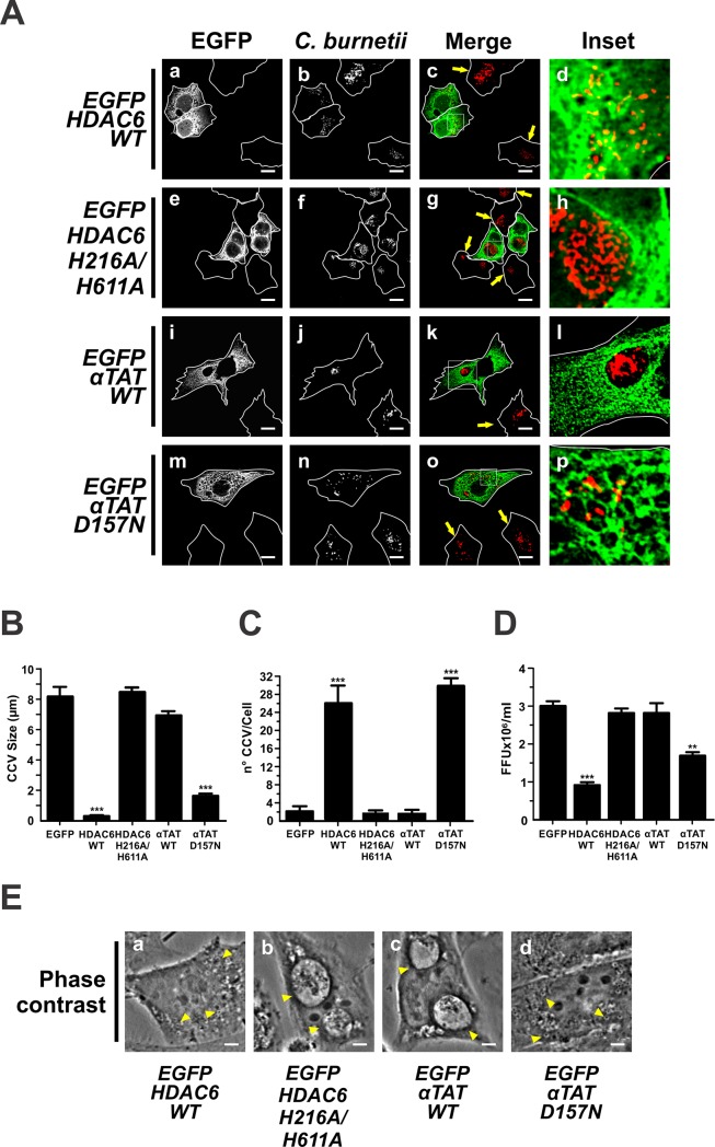
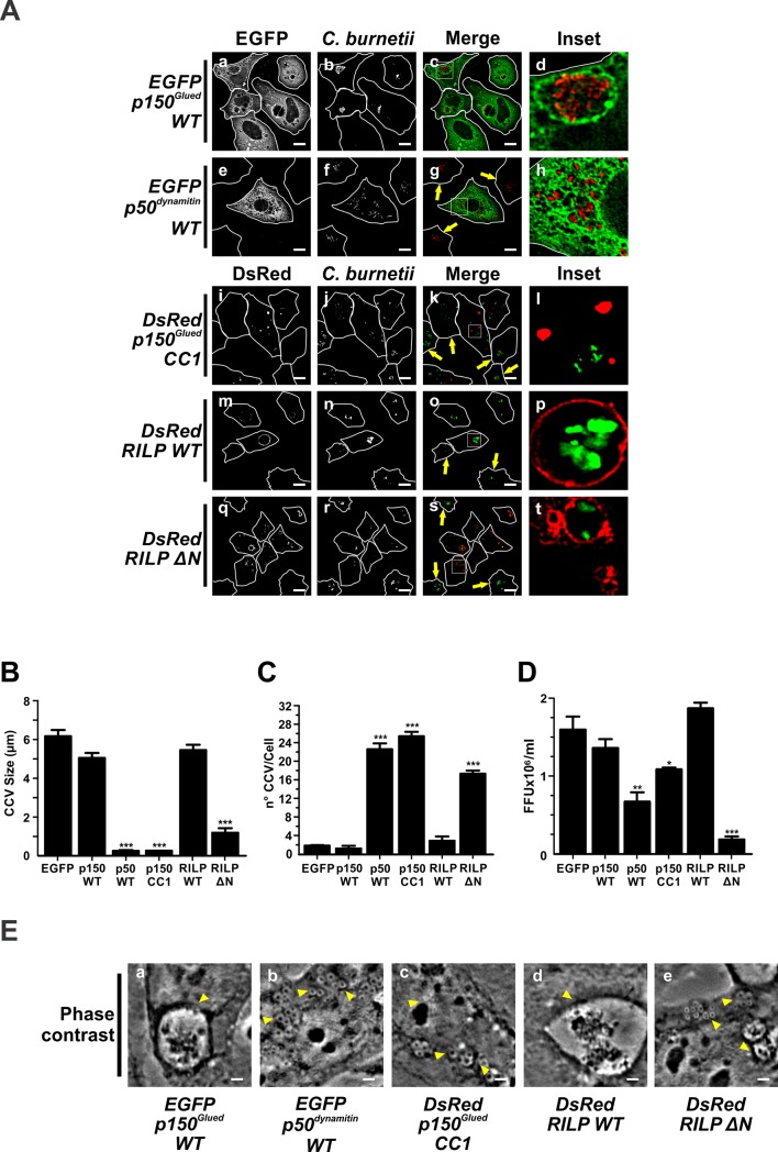
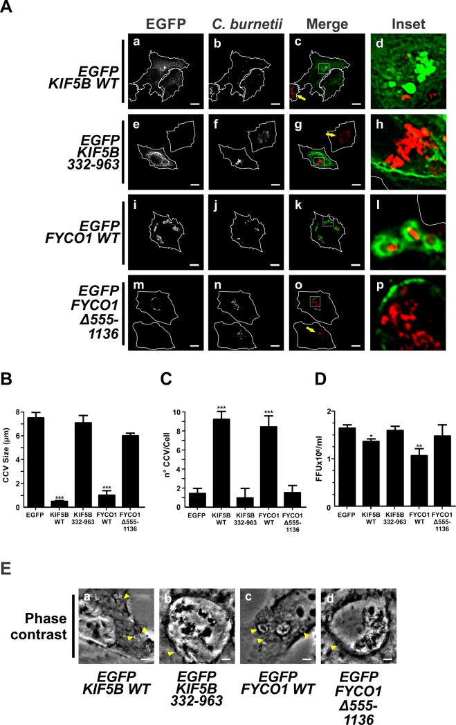
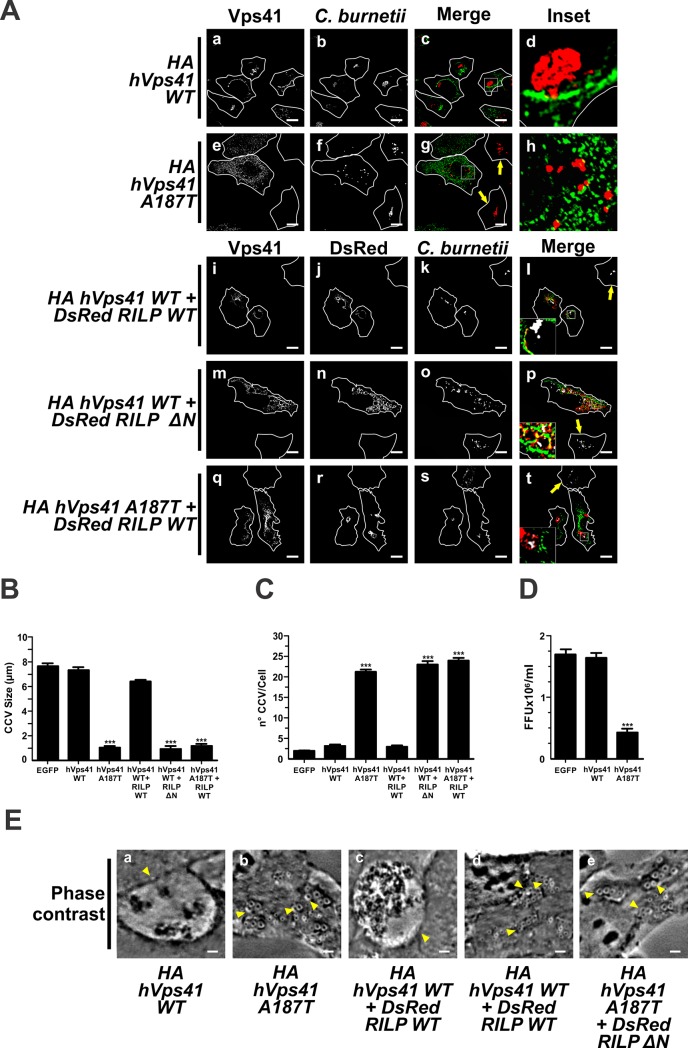
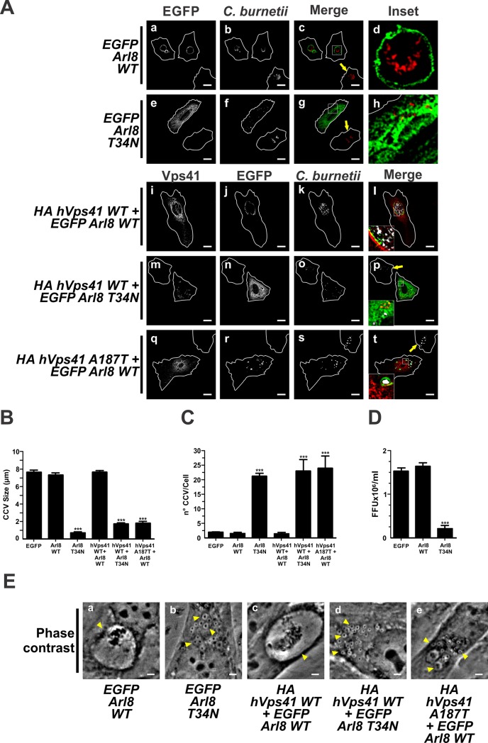
Microtubules (Mts) are dynamic cytoskeleton structures that play a key role in vesicular transport. The Mts-mediated transport depends on motor proteins named kinesins and the dynein/dynactin motor complex. The Rab7 adapter protein FYCO1 controls the anterograde transport of the endocytic compartments through the interaction with the kinesin KIF5. Rab7 and its partner RILP induce the recruitment of dynein/dynactin to late endosomes regulating its retrograde transport to the perinuclear area to fuse with lysosomes. The late endosomal-lysosomal fusion is regulated by the HOPS complex through its interaction with RILP and the GTPase Arl8. Coxiella burnetii (Cb), the causative agent of Q fever, is an obligate intracellular pathogen, which generates a large compartment with autophagolysosomal characteristics named Cb-containing vacuole (CCV). The CCV forms through homotypic fusion between small non-replicative CCVs (nrCCV) and through heterotypic fusion with other compartments, such as endosomes and lysosomes. In this work, we characterise the role of Mts, motor proteins, RILP/Rab7 and Arl8 on the CCV biogenesis. The formation of the CCV was affected when either the dynamics and/or the acetylation state of Mts were modified. Similarly, the overexpression of the dynactin subunit non-functional mutants p150Glued and RILP led to the formation of small nrCCVs. This phenomenon is not observed in cells overexpressing WT proteins, the motor KIF5 or its interacting protein FYCO1. The formation of the CCV was normal in infected cells that overexpressed Arl8 alone or together with hVps41 (a HOPS subunit) or in cells co-overexpressing hVps41 and RILP. The dominant negative mutant of Arl8 and the non-functional hVps41 inhibited the formation of the CCV. When the formation of CCV was affected, the bacterial multiplication diminished. Our results suggest that nrCCVs recruit the molecular machinery that regulate the Mts-dependent retrograde transport, Rab7/RILP and the dynein/dynactin system, as well as the tethering processes such as HOPS complex and Arl8 to finally originate the CCV where C. burnetii multiplies.


Conflict of interest statement

The authors have declared that no competing interests exist.

Figures

Fig 1
Fig 1. Dynamic microtubules participate in the formation of the CCV.
HeLa cells were infected for 16 h with Coxiella burnetii and incubated for 48 h with either DMSO (control), taxol or nocodazole. Cells were then fixed and processed for IIF. Mts were labelled with an anti-α tubulin antibody (green pseudo-colour) or C. burnetii with a specific antibody (red pseudo-colour). (A) Cells treated with 0.1% DMSO (panel a-d), 2 μM taxol (panel e-h), and 2 μM nocodazole (Noc) (panel i-l). Scale bar: 10 μm. Quantitative analysis of CCV size (B) and number (C)s, and bacterial multiplication (D). Forty to sixty cells were analysed in each experiment. Results are expressed as means ± SE of three independent experiments. ***p<0.001. (E) Phase contrast microscopy of infected and transfected HeLa cells. Arrowheads indicate a nrCCV (panel b and c), or a CCV (panel a). Scale bar: 2 μm.
Fig 2
Fig 2. Overexpression of HDAC6 and αTAT regulate the formation of CCV.
(A) Infected HeLa cells were transfected with pEGFP-HDAC6WT (panels a-d), -HDAC6 H216A/H611A (panels e-h), -αTAT WT (panels i-l) or -αTAT D157N (panels m-p). Cells were fixed and processed for IIF. An anti-C. burnetii (red pseudo-colour) antiserum was used for detecting bacteria. Arrows indicate untransfected cells containing CCVs. Scale bar: 10 μm. Quantitative analysis of CCV size (B), number (C), and bacterial multiplication (D). Forty to sixty cells were analysed in each experiment. Results are expressed as means ± SE of three independent experiments. **p<0.01, ***p<0.001. (E) Phase contrast microscopy of infected and transfected HeLa cells. Arrowheads indicate a nrCCV (panel a and d), and a CCV (panel b and c). Scale bar: 2 μm.
Fig 3
Fig 3. The formation of CCV is regulated by the dynein/dynactin motor complex.
(A) Infected HeLa cells were transfected with pEGFP-p150GluedWT (panels a-d), -p50dynamitinWT (panels e-h), pDsRed-p150GluedCC1 (panels i-l), -RILP WT (panels m-p) or -RILP ΔN (panels q-t). Arrows indicate non-transfected cells containing CCV. Scale bar: 10μm. Cells were fixed and processed for IIF. C. burnetii was detected with an anti-C. burnetii antiserum (panels b-h, red pseudo-colour; panels j-t, green pseudo-colour. Quantitative analysis of CCV size (B) and number (C), and bacterial multiplication (D). Forty to sixty cells were analysed in each experiment. Results are expressed as means ± SE of three independent experiments. *p<0.05; ***p<0.001. (E) Phase contrast microscopy of infected and transfected HeLa cells. Arrowheads indicate a nrCCV (panels b, c and e), or a CCV (panels a and d). Scale bar: 2 μm.
Fig 4
Fig 4. Rab 7 and its effector RILP are required for the formation of CCV.
(A) Infected HeLa cells were co-transfected with pDsRed-RILP WT and pEGFP-Rab7 WT (panels a-d), pDsRed-RILP ΔN and pEGFP-Rab7 WT (panels e-h), pDsRed-RILP WT and pEGFP-p150GluedWT (panels i-l), pDsRed-RILP ΔN and pEGFP-p150GluedWT (panel m-p) or pDsRed-RILP WT and pEGFP-p50dynamitinWT (panels q-t). Cells were fixed and processed for IIF. C. burnetii was detected with an anti-C. burnetii antiserum (white pseudo-colour). Arrows indicate non-transfected cells containing CCV. Scale bar: 10 μm. Quantitative analysis of CCV size (B) and number (C). Forty to sixty cells were analysed in each experiment. Results are expressed as means ± SE of three independent experiments. ***p<0.001. (D) Phase contrast microscopy of infected and transfected HeLa cells. Arrowheads indicate a nrCCV (panels b, d and e), and a CCV (panels a and c). Scale bar: 2 μm.
Fig 5
Fig 5. The overexpression of KIF5B and FYCO1 inhibits the formation of the CCV.
(A) Infected HeLa cells were transfected with EGFP-KIF5B WT (panels a-d), or -KIF5 332–963 (panels e-h) or transfected with pEGFP-FYCO1 WT (panels i-l) or -FYCO1 Δ555–1136 (panels m-p). Cells were fixed and processed for IIF. An anti-C. burnetii antiserum was used for detecting the bacteria (red pseudo colour). Arrows indicate non-transfected cell containing CCV. Scale bar: 10 μm. Quantitative analysis of CCV size (B) and number (C) and bacterial multiplication (D). Forty to sixty cells were analysed in each experiment. Results are expressed as means ± SE of three independent experiments. *p<0.05; ** p<0.01; *** p<0.001. (E) Phase contrast microscopy of infected and transfected HeLa cells. Arrowheads indicate a nrCCV (panels a and c), or a CCV (panels b and d). Scale bar: 2 μm.
Fig 6
Fig 6. HOPS and RILP participate in the development of the CCV.
(A) Infected HeLa cells were transfected with plasmids encoding HA-hVps41 WT (panels a-d) or -hVps41 A187T (panels e-h) or co-transfected with plasmids encoding HA-hVps41WT and DsRed-RILP WT (panels i-l), HA-hVps41WT and DsRed-RILPΔN (panels m-p) or HA-hVps41 A187T and DsRed-RILP WT (panel q-t). Cells were fixed and processed for IIF. C. burnetii was detected with an anti-C. burnetii antibody // antiserum (panels a-h, red pseudo-colour; panels i-t, white pseudo-colour). Arrows indicate non-transfected cells containing CCV. Scale bar: 10 μm. Quantitative analysis of CCV size (B) and number (C), and bacterial multiplication (D). Forty to sixty cells were analysed in each experiment. Results are expressed as means ± SE of three independent experiments. ***p<0.001. (E) Phase contrast microscopy of infected and transfected HeLa cells. Arrowheads indicate a nrCCV (panels b, d and e), or a CCV (panels a and c). Scale bar: 2 μm.
Fig 7
Fig 7. Arl8 and HOPS are involved in the development of the CCV.
(A) Infected HeLa cells were transfected with plasmids encoding EGFP-Arl8 WT (panels a-d) or EGFP-Arl8 T34N (panels e-h), or co-transfected with plasmids encoding HA-hVps41 WT and EGFP-Arl8 WT (panels i-l), HA-hVps41 WT and EGFP-Arl8 T34N (panels m-p) or HA-hVps41 A187T and EGFP-Arl8 WT (panels q-t). Cells were fixed and processed for IIF. C. burnetii and Vps41 were detected with an anti-C. burnetii antiserum (red pseudo-colour) and an anti-HA (green pseudo-colour) antiserum, respectively. The arrow indicates non-transfected cell containing a CCV. Scale bar: 10 μm. (B) Quantification of (B) size and (C) number of CCV and (D) bacterial multiplication. Forty to sixty cells were analysed in each experiment. Results are expressed as means ± SE of three independent experiments. ***p<0.001. (E) Phase contrast microscopy of infected and transfected HeLa cells. Arrowheads indicate a nrCCV (panels b, d and e), or a CCV (panels a and c). Scale bar: 2 μm.
Fig 8
Fig 8. Relationship of intracellular transport of C. burnetii with microtubules and motor proteins: A model.
C. burnetii (Cb) transits along the endo-phagocytic pathway into non-replicative C. burnetii-containing vacuole (nrCCVs) acquiring markers such as Rab7. This small GTPase recruits RILP protein, dynein/dynactin motor and HOPS complexes to the nrCCVs. This molecular machinery drives a gradual retrograde transport along Mts and the fusion of the nrCCVs with each other and with other endocytic compartments such as lysosomes. The fusion of lysosomes with nrCCVs is stimulated by the small GTPase Arl8 and HOPS complex. To the end of this journey, the C. burnetii-containing vacuole (CCV) is formed. Dynamic Mts and their acetylation-deacetylation status, regulated by acetyl transferase and deacetylase, are also important for CCV formation. C. burnetii could inhibit kinesin/FYCO1 (orange arrows) favouring the retrograde transport driven by the dynein/dynactin motor complex. This condition leads to the formation of the CCV. The following mechanism can explain the inhibition of the formation of the CCV by the expression of kinesin or FYCO1: the balance between dynein and kinesin recruited to nrCCVs can be shifted in favour of kinesin therefore the nrCCVs acquire a Mts-mediated anterograde movement that disperses them in the cytoplasm (not shown in the model).

References

    1. Porter SR, Czaplicki G, Mainil J, Guattéo R, Saegerman C. Q fever: Current state of knowledge and perspectives of research of a neglected zoonosis. Int J Microbiol. 2011;2011 10.1155/2011/248418 - DOI - PMC - PubMed
    1. Baca OG, Paretsky D. Q fever and Coxiella burnetii: a model for host-parasite interactions. Microbiol Rev. 1983;47: 127–149. - PMC - PubMed
    1. Hackstadt T, Peacock MG, Hitchcock PJ, Cole RL. Lipopolysaccharide variation in Coxiella burnetii: Intrastrain heterogeneity in structure and antigenicity. Infect Immun. 1985;48: 359–365. - PMC - PubMed
    1. Moos A, Hackstadt T. Comparative virulence of intra- and interstrain lipopolysaccharide variants of Coxiella burnetii in the guinea pig model. Infect Immun. 1987;55: 1144–50. - PMC - PubMed
    1. Berón W, Gutierrez MG, Rabinovitch M, Colombo MI. Coxiella burnetii localizes in a Rab7-labeled compartment with autophagic characteristics. Infect Immun. 2002;70: 5816–5821. 10.1128/IAI.70.10.5816-5821.2002 - DOI - PMC - PubMed

Publication types

MeSH terms