. 2019 Jan 14;14(1):e0210728.
doi: 10.1371/journal.pone.0210728. eCollection 2019.

Serum zonulin as a marker of intestinal mucosal barrier function: May not be what it seems

Affiliations

Serum zonulin as a marker of intestinal mucosal barrier function: May not be what it seems

Mary Ajamian et al. PLoS One. .

Abstract

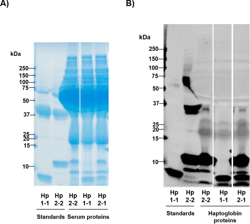
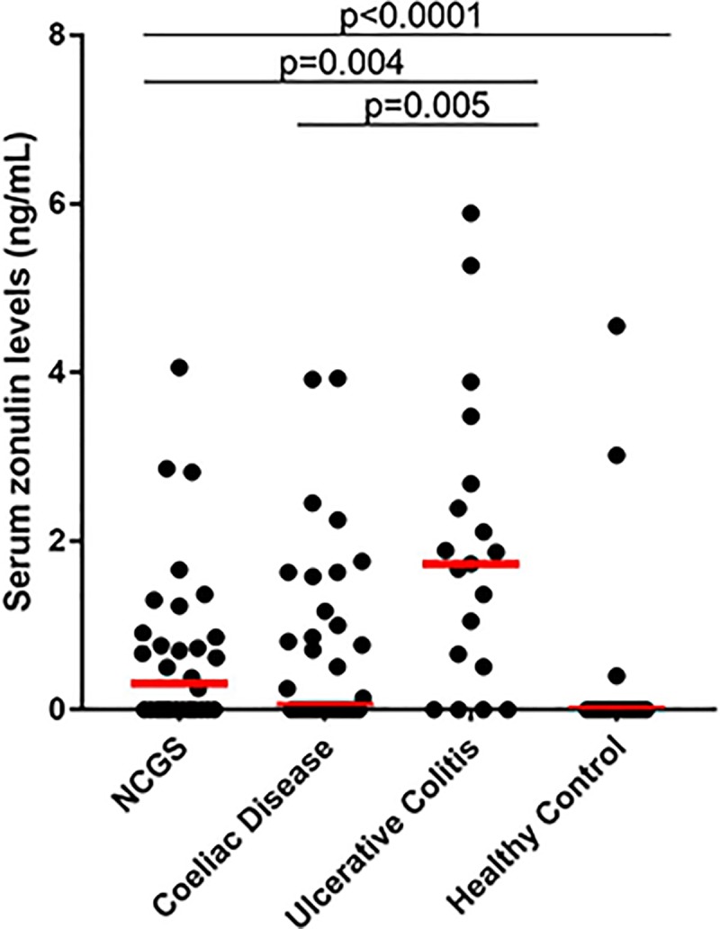
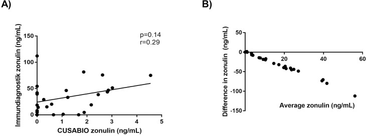
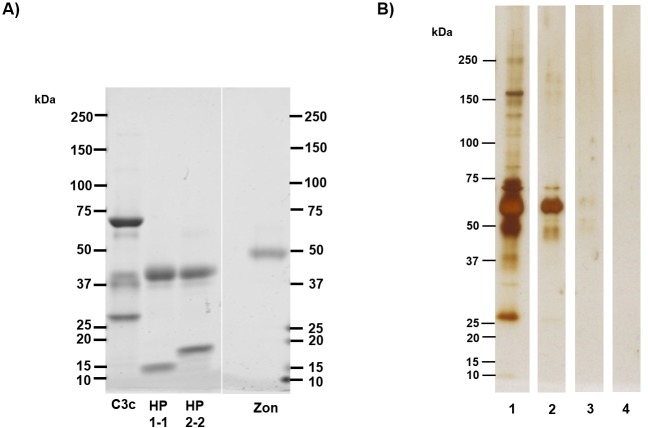
The protein, zonulin, has emerged as a popular serological marker to assess the integrity of the intestinal mucosal barrier. However, there is limited information on the utility of serum zonulin to indicate gastrointestinal disease and the validity of zonulin detection in widely-used commercial assays. The current study reports differences in zonulin levels across patient groups with gastrointestinal dysfunction compared with healthy individuals, though methodological inconsistencies indicated that actual zonulin protein was not detected by the commercial assays applied. The nature of the assays' detected antigen was investigated using immunoprecipitation followed by mass spectrometric analysis and sodium dodecyl sulphate-polyacrylamide gel electrophoresis (SDS-PAGE) followed by protein staining. Top matches of the assays' detected antigen included haptoglobin and complement C3 for the assay manufactured by CUSABIO (Wuhan, China) and complement C3 for the assay manufactured by Immundiagnostik AG (Bensheim, Germany). These findings confirm that current commercial zonulin assays are not detecting the actual protein as prehaptoglobin-2. Until assay methodology is improved, we advise the greater scientific and medical community to exercise caution in considering the measurement of serum zonulin as a marker of mucosal barrier integrity.


Conflict of interest statement

The authors have declared that no competing interests exist.

Figures

Fig 1
Fig 1. Haptoglobin phenotyping analysis by immunoblot.
Haptoglobin proteins in serum samples of patients and controls were detected by anti-haptoglobin polyclonal antibodies and compared to protein standards to determine phenotype. (A) SDS-PAGE separation followed by Coomassie-based staining of haptoglobin standards and proteins in patient sera. The first two lanes from the left contain Hp1-1 and Hp2-2 protein standards, respectively. The third, fourth, and fifth lanes from the left contain study participant sera. As expected in sera, a high abundance of albumin protein was detected in the 66.5 kDa range. (B) Immunoblot of the standards and study participant sera in which haptoglobin proteins are specifically detected by anti-haptoglobin polyclonal antibodies. The first lane from the left contains contains Hp1-1 standard, which has a characteristic band at ~9 kDa representative of the haptoglobin α1 chain. The second lane from the left contains Hp2-2 standard, with slower migration bands at higher molecular weights representative of the haptoglobin α2 chain. Hp2-2, Hp1-1, and Hp2-1 phenotypes were detected, in the third, fourth, and fifth lanes from the left, respectively.
Fig 2
Fig 2. Purported serum zonulin levels (ng/mL) in zonulin producers detected by CUSABIO ELISA assay.
Levels of zonulin, as advertised, were determined in non-coeliac wheat sensitivity (NCWS) (n = 36), coeliac disease, (n = 37), and ulcerative colitis (n = 20) patients as well as healthy individuals (n = 49). Compared with the cohort of healthy individuals, patient zonulin levels were elevated (all p<0.0001). Levels in ulcerative colitis were higher than those in NCWS (p = 0.004) and coeliac disease (p = 0.005) with no significant differences between NCWS and coeliac disease. Study samples were ran in duplicate. Red horizontal bars represent median levels for each cohort. Differences in levels between study cohorts were evaluated by Mann-Whitney U tests for nonparametric distributions.
Fig 3
Fig 3. Comparison of purported serum zonulin levels (ng/mL) between CUSABIO and Immundiagnostik ELISA assays.
Selected study samples (n = 28) ran in duplicate were compared. (A) Correlation between the two assays (p = 0.14, Spearman’s r = 0.29). (B) Bland-Altman plot calculating difference in zonulin levels vs average of zonulin levels (bias/average discrepancy between methods was -31.62 ng/mL, 95% limits of agreement were from -88.51 to 25.28 ng/mL).
Fig 4
Fig 4. Visualisation of immunoprecipitated protein products and standards.
(A) 5 μg of human complement C3c, HP1-1, HP2-2 and recombinant zonulin (zon) standards were separated by SDS-PAGE and stained with Coomassie gel stain. Characteristic bands of standards include the conserved C3c β-chain at 70 kDa, the haptoglobin β-chain at 40 kDa, and the 47 kDa band indicative of recombinant zonulin. (B) Silver staining of immunoprecipitated protein samples detected by LC-MS/MS. Lanes (1) and (2) contain immunoprecipitated proteins from incubation with serum in commercial assays. Lane (1) contains serum proteins captured by CUSABIO assay, which includes complement C3 and haptoglobin as identified by mass spectrometry. Bands at 70 kDa, suggestive of complement C3, and haptoglobin at 40 kDa are present. Lane (2) contains serum proteins captured by Immundiagnostik assay, which includes complement C3 as identified by mass spectrometry. Lanes (3) and (4) contain immunoprecipitated product from incubation of commercial kits with recombinant zonulin. Lane (3), which includes immunoprecipitated product captured by CUSABIO assay, contains trace amounts of protein around the 45–65 kDa range. Mass spectrometry results also indicated a small amount of haptoglobin (S1 File), which remained undetected by silver stain. Lane (4) contained immunoprecipitated product captured by Immundiagnostik assay; no significant detectable proteins aside from albumin were detected by mass spectrometry (S1 File) or indicated by silver stain.

References

    1. Odenwald MA, Turner JR. Intestinal permeability defects: is it time to treat? Clinical gastroenterology and hepatology: the official clinical practice journal of the American Gastroenterological Association. 2013;11(9):1075–83. 10.1016/j.cgh.2013.07.001 - DOI - PMC - PubMed
    1. Fasano A. Zonulin and its regulation of intestinal barrier function: the biological door to inflammation, autoimmunity, and cancer. Physiological reviews. 2011;91(1):151–75. 10.1152/physrev.00003.2008 . - DOI - PubMed
    1. Turner JR. Intestinal mucosal barrier function in health and disease. Nature reviews Immunology. 2009;9(11):799–809. 10.1038/nri2653 . - DOI - PubMed
    1. Fasano A, Shea-Donohue T. Mechanisms of disease: the role of intestinal barrier function in the pathogenesis of gastrointestinal autoimmune diseases. Nature clinical practice Gastroenterology & hepatology. 2005;2(9):416–22. 10.1038/ncpgasthep0259 . - DOI - PubMed
    1. Tripathi A, Lammers KM, Goldblum S, Shea-Donohue T, Netzel-Arnett S, Buzza MS, et al. Identification of human zonulin, a physiological modulator of tight junctions, as prehaptoglobin-2. Proceedings of the National Academy of Sciences of the United States of America. 2009;106(39):16799–804. 10.1073/pnas.0906773106 - DOI - PMC - PubMed

Publication types