. 2019 Jan 11;20(2):270.
doi: 10.3390/ijms20020270.

IL-1β Inflammatory Cytokine-Induced TP63 Isoform ∆NP63α Signaling Cascade Contributes to Cisplatin Resistance in Human Breast Cancer Cells

Affiliations

IL-1β Inflammatory Cytokine-Induced TP63 Isoform ∆NP63α Signaling Cascade Contributes to Cisplatin Resistance in Human Breast Cancer Cells

Mónica G Mendoza-Rodríguez et al. Int J Mol Sci. .

Abstract

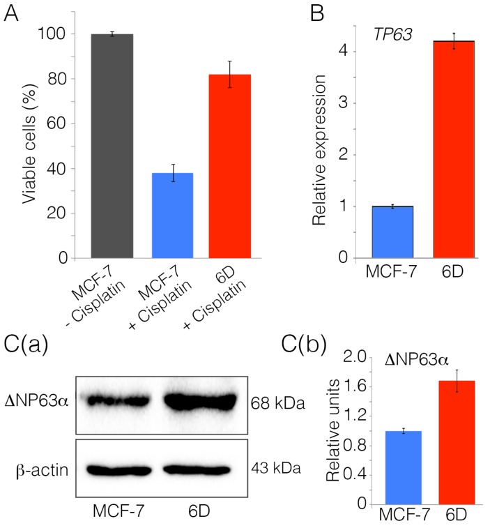
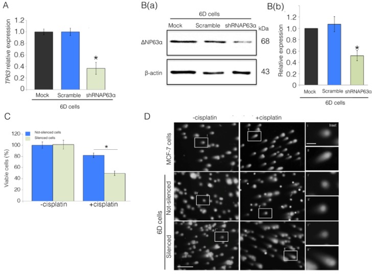
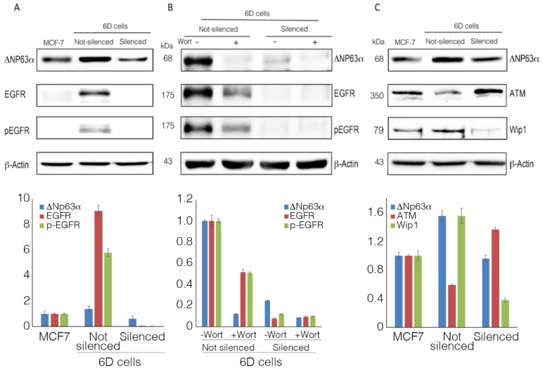
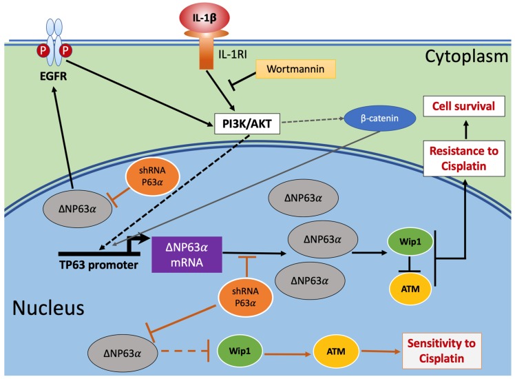
The mechanisms behind the induction of malignancy and chemoresistance in breast cancer cells are still not completely understood. Inflammation is associated with the induction of malignancy in different types of cancer and is highlighted as an important factor for chemoresistance. In previous work, we demonstrated that the inflammatory cytokine interleukin 1β (IL-1β)-induced upregulation of genes was associated with chemoresistance in breast cancer cells. Here, we evaluated the participation and the expression profile of TP63 in the induction of resistance to cisplatin. By loss-of-function assays, we identified that IL-1β particularly upregulates the expression of the tumor protein 63 (TP63) isoform ΔNP63α, through the activation of the IL-1β/IL-1RI/β-catenin signaling pathway. Upregulation of ΔNP63α leads to an increase in the expression of the cell survival factors epidermal growth factor receptor (EGFR) and phosphatase 1D (Wip1), and a decrease in the DNA damage sensor, ataxia-telangiectasia mutated (ATM). The participation of these processes in the increase of resistance to cisplatin was confirmed by silencing TP63 expression or by inhibition of the phosphoinositide 3-kinase (PI3K)/protein kinase B (AKT) activity in the IL-1β/IL-1RI/β-catenin signaling pathway. These data reinforced the importance of an inflammatory environment in the induction of drug resistance in cancer cells and uncovered a molecular mechanism where the IL-1β signaling pathway potentiates the acquisition of cisplatin resistance in breast cancer cells.

Keywords: IL-1β; TP63 isoform ΔNP63α; breast cancer; drug resistance acquisition; short hairpin RNA (shRNA)-mediated knockdown.


Conflict of interest statement

The authors declare no conflicts of interest.

Figures

Figure 1
Figure 1
Interleukin 1β (IL-1β) induces resistance to cisplatin and upregulates the expression of TP63. (A) Non-invasive MCF-7 cells and invasive 6D cells were treated with 100 µM cisplatin (MCF-7 + cisplatin) and (6D + cisplatin). MCF-7 cells without any treatment were utilized as the control of live cells in the assay (100% viability). Cells were harvested at 48 h and quantification of viable cells performed using the WST-1 reagent. Data are presented as percentage ± SD of viable cells relative to untreated cells from three independent experiments. (B) Relative expression of the TP63 gene was determined by qPCR in MCF-7 and 6D cells. Results represent the average of three independent experiments ± SD. (C) (a,b) Representative Western blot and densitometry analysis of total extracts from MCF-7 and 6D cells. The membranes were challenged with anti-TP63 antibody and anti-β-actin for protein load control. The densitometric analysis shows data in three blots from independent experiments. In all of them, ΔNp63α levels were normalized relative to the protein levels in MCF-7 cells. Asterisks indicate significance at p = 0.001.
Figure 2
Figure 2
ΔNp63α plays a role in the IL-1β induction of cisplatin resistance in 6D cells. Cells were transfected with empty vector (Mock), non-specific short hairpin RNA (Scramble), and the specific silencing RNA (shRNAp63α). (A) TP63 expression was evaluated by qPCR. Results represent the average of three independent experiments ± SD. Asterisks correspond to p = 0.001 relative to the controls, Mock and Scramble. (B(a)) Representative Western blot of ΔNp63α protein levels in the 6D cells. (B(b)) Densitometric values corresponding to ΔNp63α levels in (B(a)) were normalized to those of β-actin. (C) Cell viability levels determined in ΔNp63α-silenced and non-silenced cells in the absence or presence of cisplatin. Data represent the average of four independent experiments ± SD. Asterisks indicate significance relative to 6D cells at p = 0.001. (D) Comet assay to evaluate DNA integrity and damage by cisplatin in MCF-7, non-silenced, and shRNAp63α-silenced 6D cells. Cells not treated and treated with cisplatin were mixed with low-melting point agarose, lysed, and subjected to electrophoresis, followed by staining with ethidium bromide. DNA was visualized by fluorescence microscopy; scale bar = 100 μm. The insets in the right panel show magnified images taken from each condition; scale bar = 25 μm.
Figure 3
Figure 3
ΔNp63α is essential for the expression of epidermal growth factor receptor (EGFR) and phosphatase 1D (Wip1). (A) Representative Western blot of EGFR expression and its activated form pEGFR (phosphorylated at Tyr1068) in parental MCF-7cells and in non-silenced and shRNAp63α-silenced 6D cells. (B) Wortmannin’s inhibitory effect on ΔNp63α and EGFR overexpression and phosphorylation of the latter. (C) Western blot analysis of ataxia-telangiectasia mutated (ATM) and Wip1 expression in the cells indicated in panel A. Densitometric analyses of Western blots show the results of at least three independent experiments.
Figure 4
Figure 4
Graphical model of IL-1β activation of signaling pathways leading to acquisition of resistance to cisplatin in breast cancer cells. Triggering of the IL-1β/IL-1RI/β-catenin pathway by IL-1β induces the activity of downstream effectors phosphoinositide 3-kinase (PI3K)/protein kinase B (AKT) [4], leading to translocation of β-catenin to the nucleus and regulation of proteins that participate in the acquisition of resistance to cisplatin. The induced upregulation of ΔNp63α regulates the expression of cell survival (pEFGR) and DNA damage response (Wip1 and ATM) proteins. At the same time, the increased levels of ΔNP63α activate EGFR, which, through a feed-back loop, maintains PI3K/AKT activity and the continuity of the downstream signaling initiated by IL-1β, which leads to resistance to cisplatin. However, if ΔNP63α overexpression is hampered by silencing its mRNA (shRNAP63α), the lower levels of ΔNP63α expressed in the cells will not be sufficient to maintain high levels of Wip1, thereby increasing ATM levels and the sensitivity to cisplatin. Dashed arrows indicate indirect positive modulation in the pathway that was previously reported [4,14]. The link between low levels of ΔNp63α and Wip1 is represented in the figure with a dashed line because the step(s) leading to decreased levels of Wip1 are not yet well identified.

References

    1. Zitvogel L., Kepp O., Galluzzi L., Kroemer G. Inflammasomes in carcinogenesis and anticancer immune responses. Nat. Immunol. 2012;13:343–351. doi: 10.1038/ni.2224. - DOI - PubMed
    1. Multhoff G., Molls M., Radons J. Chronic inflammation in cancer development. Front. Immunol. 2011;2:98. doi: 10.3389/fimmu.2011.00098. - DOI - PMC - PubMed
    1. Perez-Yepez E.A., Ayala-Sumuano J.T., Reveles-Espinoza A.M., Meza I. Selection of a MCF-7 Breast Cancer Cell Subpopulation with High Sensitivity to IL-1beta: Characterization of and Correlation between Morphological and Molecular Changes Leading to Increased Invasiveness. Int. J. Breast Cancer. 2012;2012:609148. doi: 10.1155/2012/609148. - DOI - PMC - PubMed
    1. Perez-Yepez E.A., Ayala-Sumuano J.T., Lezama R., Meza I. A novel beta-catenin signaling pathway activated by IL-1beta leads to the onset of epithelial-mesenchymal transition in breast cancer cells. Cancer Lett. 2014;354:164–171. doi: 10.1016/j.canlet.2014.08.015. - DOI - PubMed
    1. Mendoza-Rodriguez M., Arevalo Romero H., Fuentes-Panana E.M., Ayala-Sumuano J.T., Meza I. IL-1beta induces up-regulation of BIRC3, a gene involved in chemoresistance to doxorubicin in breast cancer cells. Cancer Lett. 2017;390:39–44. doi: 10.1016/j.canlet.2017.01.005. - DOI - PubMed