Review
. 2019 Jan 11;20(2):280.
doi: 10.3390/ijms20020280.

Bone Metastasis Pain, from the Bench to the Bedside

Affiliations
Review

Bone Metastasis Pain, from the Bench to the Bedside

Federica Aielli et al. Int J Mol Sci. .

Abstract

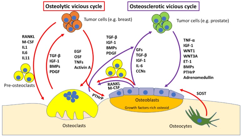
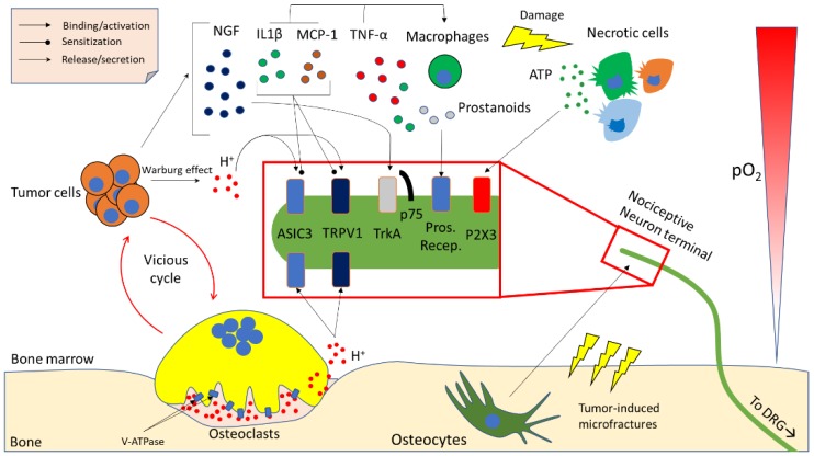
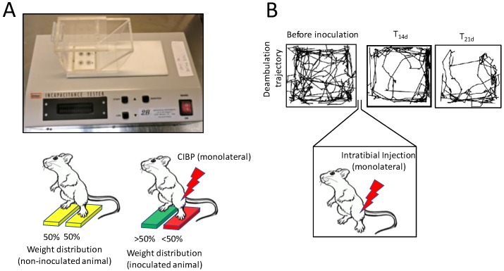
Bone is the most frequent site of metastasis of the most common cancers in men and women. Bone metastasis incidence has been steadily increasing over the years, mainly because of higher life expectancy in oncologic patients. Although bone metastases are sometimes asymptomatic, their consequences are most often devastating, impairing both life quality and expectancy, due to the occurrence of the skeletal-related events, including bone fractures, hypercalcemia and spinal cord compression. Up to 75% of patients endure crippling cancer-induced bone pain (CIBP), against which we have very few weapons. This review's purpose is to discuss the molecular and cellular mechanisms that lead to CIBP, including how cancer cells convert the bone "virtuous cycle" into a cancer-fuelling "vicious cycle", and how this leads to the release of molecular mediators of pain, including protons, neurotrophins, interleukins, chemokines and ATP. Preclinical tests and assays to evaluate CIBP, including the incapacitance tester (in vivo), and neuron/glial activation in the dorsal root ganglia/spinal cord (ex vivo) will also be presented. Furthermore, current therapeutic options for CIBP are quite limited and nonspecific and they will also be discussed, along with up-and-coming options that may render CIBP easier to treat and let patients forget they are patients.

Keywords: bone metastasis; bone pain; osteoblasts; osteoclasts; skeletal-related events.


Conflict of interest statement

The authors declare no conflict of interest.

Figures

Figure 1
Figure 1
Cartoon representing the osteolytic and osteosclerotic vicious cycles. Osteolytic cycle: osteolysis-generating cancer cells (e.g., breast) migrate to the bone marrow and start secreting osteoclast-stimulating factors such as epithelial growth factor (EGF), osteoclast stimulating factor (OSF), tumour necrosis factors (TNFs) and Activin A. In parallel, cancer cells also secrete osteoclast-differentiating factors, such as receptor activator of nuclear factor kB ligand (RANKL), macrophage-colony stimulating factor (M-CSF) and Interleukin (IL)-1, 6 and 11, which promote differentiation of pre-osteoclasts into osteoclasts. The bone matrix is rich in growth factors (GFs), transforming growth factor (TGF)-β, insulin-like growth factor (IGF)-1, bone morphogenic proteins (BMPs) and platelet-derived growth factor (PDGF). These factors get released when osteoclasts resorb bone, and they promote tumour growth, which closes the osteolytic vicious cycle. Osteocytes also take a crucial part in the process, secreting sclerostin (SOST) in response to osteolytic cancers (especially multiple myeloma), which inhibits osteoblast activity and the Wnt- β-catenin pathway Osteosclerotic cycle: osteosclerosis-generating cancer cells (e.g., prostate) migrate to the bone marrow and start secreting osteoblast-stimulating factors such as TNF-α, IGF-1, Wingless-type MMTV integration site (WNT)1, WNT3A, Endothelin (ET)-1, BMPs, parathyroid hormone-related peptide (PTHrP) and adrenomedullin. This stimulates osteoblast differentiation and activity. On one hand this leads to osteoblast-mediated osteoclastogenesis by increasing osteoblastic expression of RANKL and M-CSF, which causes the release of growth factors as discussed for the osteolytic cycle. On the other hand, osteoblasts themselves release a plethora of factors including GFs, TGF-β, IGF-1, IL-6 and chemokines (CCNs), which stimulate tumour growth, closing the osteosclerotic (which most of the times has an osteolytic component as well) vicious cycle.
Figure 2
Figure 2
Cartoon representing the main cellular and molecular players in cancer-induced bone pain (CIBP). Partial pressure of O2 (pO2) decreases to approximately 50 mmHg moving from bone marrow sinusoids towards bone. When tumour cells are present in the bone marrow microenvironment, the low pO2, along with other factors, causes the switch from a mostly oxidative to a mostly glycolytic glucose metabolism, which results in the production of lactate and protons (H+) that are released in the microenvironment to keep intracellular pH stable (Warburg effect). Cancer cells also promote osteoclast formation and activity. Osteoclasts release protons into the resorption lacuna through the vacuolar-ATPase (V-ATPase). Protons may then leak towards the bone marrow microenvironment due to improper seal or apoptosis of the osteoclast. Protons are subsequently able to activate acid sensing ion channels (ASIC)-3, and transient receptor potential channel-vanilloid subfamily members (TRPV)-1, on nociceptive neuron terminals arising from dorsal root ganglia (DRGs), which causes the activation of pain pathways. Osteocytes can activate nociceptive neurons in different ways in response to tumour-induced microfractures, but they are mainly thought to release protons as signalling molecule, although other molecules such as neurotrophins have been proposed as osteocyte-derived pain-mediating factors. Tumour cells also secrete many factors that are able to activate or sensitise pain-mediating receptors, such as nerve growth factor (NGF), which binds the tropomyosin-related kinase (Trk)A-p75 receptor complex. Also, tumour-derived interleukin (IL)-1β, along with macrophage chemoattractant protein (MCP)-1 can sensitise ASIC3 and TRPV1 and activate and chemoattract macrophages. These latter cells further secrete IL-1β, MCP-1 and tumour necrosis factor (TNF)-α, which act as autocrine factors as well as induce cell death in the tissue. Macrophages also produce prostanoids, which activate nociception through prostanoid receptors. Because of the strong immune response, reactive oxygen species (ROS) generation, cancer-induced cytotoxicity and many other factors, cells in the bone marrow microenvironment undergo necrosis, which leads to the release of ATP. ATP can then act as a pain mediator by activating the purinergic P2X3 receptor on nociceptive neuron terminal.
Figure 3
Figure 3
In vivo, noninvasive methods to evaluate cancer-induced bone pain (CIBP). (A) Incapacitance tester: cancer cells are intratibially injected monolaterally, and after an experiment-specific timeframe the animal is placed on an incapacitance tester. This is a device which features 2 scales, that are able to discriminate weight distribution between the 2 hindlimbs, when the animal is stood up at an incline (as visible from A, upper panel). In normal conditions, rodents will tend to distribute the weight evenly between the 2 limbs, but when one of them experiences CIBP, mice will relieve them from some of their body weight, reducing the % of weight bore by that limb. (B) Spontaneous deambulation test: mice are acclimated in a 45 × 45 × 45 cm arena 3 times the week before the cancer cells inoculation to establish a baseline. On the third test, the trajectory of the mouse is recorded and quantified over a specific timeframe (e.g., 10 min), to assess the distance the mouse is willing to walk voluntarily, without external stimulation. Cancer cells are then injected monolaterally (e.g., intratibially) and after an appropriate time (e.g., T14 and T21 days), mice will start showing a decrease in spontaneous ambulation, which is mostly due to CIBP. It is also possible to review the mouse behaviour to assess rearing behaviour and limb usage if a video recording device is used for the test.

References

    1. Rucci N., Teti A. The “love-hate” relationship between osteoclasts and bone matrix. Matrix Biol. 2016;54:176–190. doi: 10.1016/j.matbio.2016.02.009. - DOI - PubMed
    1. Capulli M., Paone R., Rucci N. Osteoblast and osteocyte: Games without frontiers. Arch. Biochem. Biophys. 2014;561:3–12. doi: 10.1016/j.abb.2014.05.003. - DOI - PubMed
    1. Cappariello A., Ponzetti M., Rucci N. The “soft” side of the bone: Unveiling its endocrine functions. Horm. Biol. Clin. Investig. 2016;28:5–20. doi: 10.1515/hmbci-2016-0009. - DOI - PubMed
    1. Jähn K., Kelkar S., Zhao H., Xie Y., Tiede-Lewis L.M., Dusevich V., Dallas S.L., Bonewald L.F. Osteocytes acidify their microenvironment in response to PTHrP in vitro and in lactating mice in vivo. J. Bone Miner. Res. 2017;32:1761–1772. doi: 10.1002/jbmr.3167. - DOI - PMC - PubMed
    1. Uda Y., Azab E., Sun N., Shi C., Pajevic P.D. Osteocyte mechanobiology. Curr. Osteoporos. Rep. 2017;15:318–325. doi: 10.1007/s11914-017-0373-0. - DOI - PMC - PubMed