

Pulmonary exposure to peat smoke extracts in rats decreases expiratory time and increases left heart end systolic volume
- PMID: 30642191
- PMCID: PMC7319252
- DOI: 10.1080/08958378.2018.1551443
Pulmonary exposure to peat smoke extracts in rats decreases expiratory time and increases left heart end systolic volume
Abstract
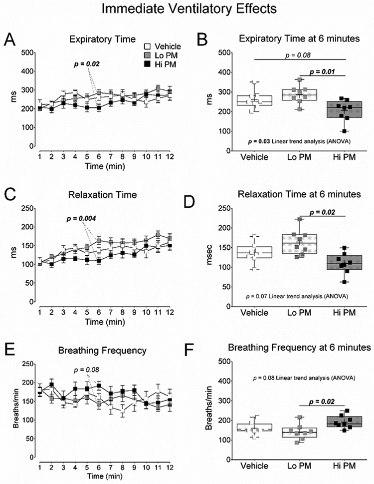
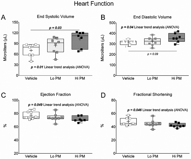
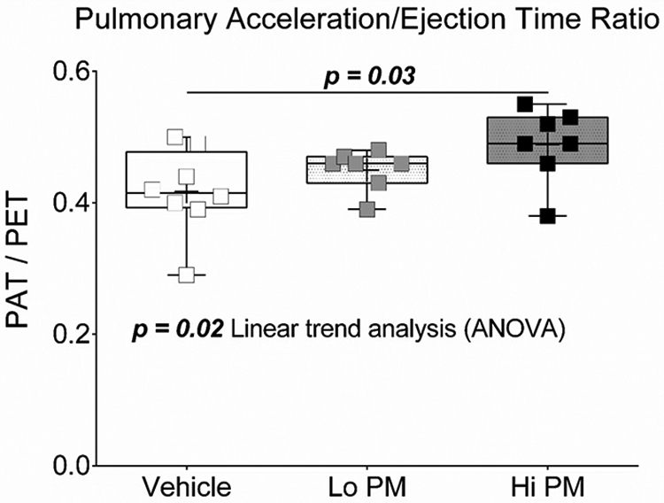
Exposure to wildland fire-related particulate matter (PM) causes adverse health outcomes. However, the impacts of specific biomass sources remain unclear. The purpose of this study was to investigate cardiopulmonary responses in rats following exposure to PM extracts collected from peat fire smoke. We hypothesized that peat smoke PM would dose-dependently alter cardiopulmonary function. Male Sprague-Dawley rats (n = 8/group) were exposed to 35 µg (Lo PM) or 350 µg (Hi PM) of peat smoke PM extracts suspended in saline, or saline alone (Vehicle) via oropharyngeal aspiration (OA). Ventilatory expiration times, measured in whole-body plethysmographs immediately after OA, were the lowest in Hi PM exposed subjects at 6 min into recovery (p = .01 vs. Lo PM, p = .08 vs. Vehicle) and resolved shortly afterwards. The next day, we evaluated cardiovascular function in the same subjects via cardiac ultrasound under isoflurane anesthesia. Compared to Vehicle, Hi PM had 45% higher end systolic volume (p = .03) and 17% higher pulmonary artery blood flow acceleration/ejection time ratios, and both endpoints expressed significant increasing linear trends by dose (p = .01 and .02, respectively). In addition, linear trend analyses across doses detected an increase for end diastolic volume and decreases for ejection fraction and fractional shortening. These data suggest that exposure to peat smoke constituents modulates regulation of ventricular ejection and filling volumes, which could be related to altered blood flow in the pulmonary circulation. Moreover, early pulmonary responses to peat smoke PM point to irritant/autonomic mechanisms as potential drivers of later cardiovascular responses.
Keywords: Wildfire; biomass; echocardiography; heart function; particulate matter; peat; pulmonary irritation; smoke; ultrasound; wildland fire.
Figures
References
-
- Argacha JF, Bourdrel T, van de Borne P. 2017. Ecology of the cardiovascular system: A focus on air-related environmental factors. Trends Cardiovasc Med. - PubMed
-
- Baker KR, Woody MC, Valin L, Szykman J, Yates EL, Iraci LT, Choi HD, Soja AJ, Koplitz SN, Zhou L et al. 2018. Photochemical model evaluation of 2013 California wild fire air quality impacts using surface, aircraft, and satellite data. Sci Total Environ. 637-638:1137–1149. eng. - PubMed
-
- Brook RD, Rajagopalan S, Pope CA 3rd, Brook JR, Bhatnagar A, Diez-Roux AV, Holguin F, Hong Y, Luepker RV, Mittleman MA et al. 2010. Particulate matter air pollution and cardiovascular disease: An update to the scientific statement from the American Heart Association. Circulation. 121(21):2331–2378. - PubMed
-
- Carll AP, Haykal-Coates N, Winsett DW, Hazari MS, Ledbetter AD, Richards JH, Cascio WE, Costa DL, Farraj AK. 2015. Cardiomyopathy confers susceptibility to particulate matter-induced oxidative stress, vagal dominance, arrhythmia and pulmonary inflammation in heart failure-prone rats. Inhalation toxicology. 27(2):100–112. - PMC - PubMed
MeSH terms
Substances
Grants and funding
LinkOut - more resources
Full Text Sources