

Identifying lesions in paediatric epilepsy using morphometric and textural analysis of magnetic resonance images
- PMID: 30642755
- PMCID: PMC6412079
- DOI: 10.1016/j.nicl.2019.101663
Identifying lesions in paediatric epilepsy using morphometric and textural analysis of magnetic resonance images
Abstract
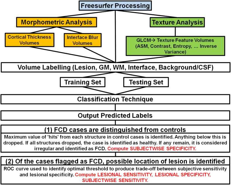
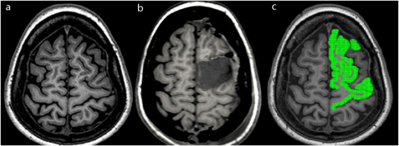
We seek to examine the use of an image processing pipeline on Magnetic Resonance Imaging (MRI) to identify features of Focal Cortical Dysplasia (FCD) in children who were suspected to have FCD on MRI (MRI-positive) and those with MRI-negative epilepsy. We aim to use a computer-aided diagnosis system to identify epileptogenic lesions with a combination of established morphometric features and textural analysis using Gray-Level Co-occurrence Matrices (GLCM) on MRI sequences. We implemented a modified version of the 2-Step Bayesian classifier method to a paediatric cohort with medically intractable epilepsy with MRI-positive and MRI-negative epilepsy, and evaluated the performance of the algorithm trained on textural features derived from T1-weighted (T1-w), T2-weighted (T2-w), and FLAIR (Fluid Attenuated Inversion Recovery) sequences. For MRI-positive cases, T1-w has the highest subjectwise sensitivity relative to T2-w and FLAIR (94% vs. 90% vs. 71% respectively), and also the highest lesional sensitivity (63% vs. 60% vs. 42% respectively), but the lowest lesional specificity (75% vs. 80% vs. 89% respectively). Combination of all three sequences improved the performance of the algorithm, with 97% subjectwise sensitivity. For MRI-negative cases, T1-w has the highest subjectwise sensitivity relative to T2-w and FLAIR (48% vs. 30% vs. 39% respectively), and also the highest lesional sensitivity (31% vs. 22% vs. 28% respectively). However, T2-w has the highest lesional specificity relative to T1-w and FLAIR (95% vs. 94% vs. 92% respectively) for MRI-negative cases. Combination of all three sequences improved the performance of the algorithm, with 70% subjectwise sensitivity. The 2-Step Naïve Bayes classifier correctly rejected 100% of the healthy subjects for all three sequences. Using combined morphometric and textural analysis in a 2-Step Bayesian classifier, applied to multiple MRI sequences, can assist with lesion detection in children with intractable epilepsy.
Keywords: Computer-aided diagnosis; Epilepsy; Focal cortical dysplasia; Morphometric and textural analysis.
Copyright © 2019 The Authors. Published by Elsevier Inc. All rights reserved.
Figures
References
-
- Alshafai L., Ochi A., Go C., McCoy B., Hawkins C., Otsubo H.…Widjaja E. Clinical, EEG, MRI, MEG, and surgical outcomes of pediatric epilepsy with astrocytic inclusions versus focal cortical dysplasia. Epilepsia. 2014;55(10):1568–1575. - PubMed
-
- Antel S.B., Collins D.L., Bernasconi N., Andermann F., Shinghal R., Kearney R.E.…Bernasconi A. Automated detection of focal cortical dysplasia lesions using computational models of their MRI characteristics and texture analysis. NeuroImage. 2003;19(4):1748–1759. - PubMed
-
- Dale A.M., Sereno M.I. Improved localizadon of cortical activity by combining EEG and MEG with MRI cortical surface reconstruction: a linear approach. J. Cogn. Neurosci. 1993;5(2):162–176. - PubMed
