Review
. 2019 May;27(5):398-407.
doi: 10.1016/j.tim.2018.12.013. Epub 2019 Jan 11.

Interplay between Influenza Virus and the Host RNA Polymerase II Transcriptional Machinery

Affiliations
Review

Interplay between Influenza Virus and the Host RNA Polymerase II Transcriptional Machinery

Alexander P Walker et al. Trends Microbiol. 2019 May.

Abstract

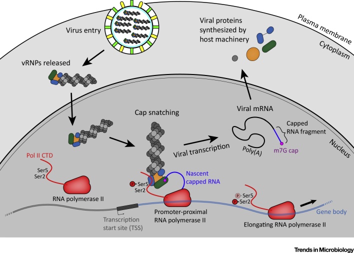
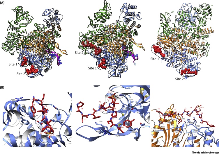
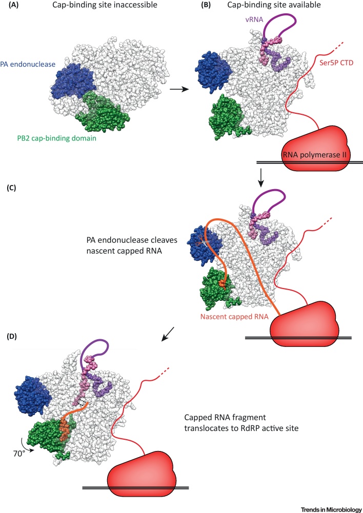
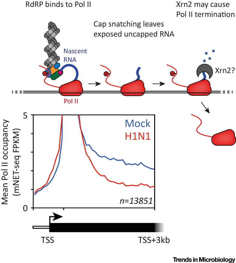
The influenza virus RNA-dependent RNA polymerase (RdRP) cleaves the 5' end of nascent capped host RNAs and uses the capped RNA fragment to prime viral transcription in a mechanism called 'cap snatching'. Cap snatching requires an intimate association between influenza RdRP and cellular RNA polymerase II (Pol II), which is the source of nascent capped host RNAs targeted by influenza virus. Recent structural studies have revealed how influenza RdRP binds to Pol II and how this binding promotes the initiation of viral transcription by influenza RdRP. In this review we focus on these recent insights into the mechanism of cap snatching by influenza virus and the impact of cap snatching on host gene expression during influenza virus infection.

Keywords: CTD; Pol II; RNA polymerase; cap snatching; influenza virus.


Figures

Figure 1
Figure 1
The Association of Influenza vRNPs with Cellular Pol II. Influenza virions bind to receptors at the plasma membrane and are endocytosed. Viral and endosomal membranes fuse, releasing vRNPs into the cytoplasm. vRNPs are then trafficked to the nucleus to target cellular Pol II. The CTD of unengaged Pol II is hypophosphorylated at Ser2 and Ser5. Pol II initiates at the transcription start site, and is Ser5-phosphorylated early in transcription. Later in transcription elongation, Ser2 is phosphorylated and Ser5 is gradually dephosphorylated. Influenza vRNPs in the nucleus target the Ser5-phosphorylated Pol II CTD, and bind to the m7G cap of nascent RNA on Pol II. RdRP in the vRNP cap snatches the nascent RNA, which is then used to prime viral transcription by influenza RdRP. The m7G capped, polyadenylated viral mRNA is exported to the cytoplasm through host pathways, and translated by host machinery.
Figure 2
Figure 2
Structures of Influenza RdRP Pol II CTD Binding Sites. (A) Ribbon structures of FluA RdRP (left) (PDB:5M3H), FluB RdRP (centre) (PDB:5M3J), and FluC RdRP (right) (PDB:6F5P) bound to Pol II CTD peptides. The PB1 subunit is coloured orange, PB2 subunit green, and PA subunit blue. CTD peptides are shown as red spheres, or mesh for the unresolved density on FluB RdRP. (B) Detailed views of CTD Site 1 (left) and Site 2 (centre) on FluA RdRP, and Site 1 on FluC RdRP (right).
Figure 3
Figure 3
Influenza RdRP Conformational Rearrangements during Cap Snatching. (A) In the transcriptionally inactive RdRP conformation, the PB2 cap-binding domain (green) is inaccessible and cannot bind capped RNA (PDB:5D98). (B) When influenza RdRP binds to vRNA and Ser5P Pol II CTD the transcriptionally active conformation is favoured. In this conformation the PB2 cap-binding domain is accessible, and is orientated towards the PA subunit endonuclease domain (blue) (PDB:4WSB). (C) Influenza RdRP binds to nascent capped RNA (orange), then the PA endonuclease cleaves 10–13 nt downstream of the m7G cap (PDB:6EVK). (D) Following cleavage, the PB2 cap-binding domain rotates by 70 degrees. This moves the capped RNA fragment away from the PA endonuclease and into the RdRP active site through the product exit channel. The capped RNA fragment can then base pair with the 3′ end of the vRNA template (pink) in the active site (PDB:5MSG).
Figure 4
Figure 4
Pol II Termination as a Result of Cap Snatching. Sequencing data (bottom) show that Pol II occupancy, measured in mNET-Seq ‘fragments per kilobase per million mapped reads’, is decreased in gene bodies during influenza virus infection (reproduced from Bauer et al. (2018) under the Creative Commons Attribution License (CC BY)). This indicates that influenza virus infection induces premature Pol II termination. In the proposed model (top), cap snatching by influenza RdRP leaves uncapped RNA with an exposed 5′ monophosphate emerging from the Pol II active site. This RNA is a substrate for Xrn2, a host exoribonuclease, which induces Pol II termination according to the torpedo model. TSS indicates transciption start site.

References

    1. Krammer F. Influenza. Nat. Rev. Dis. Prim. 2018;4:1–21. - PMC - PubMed
    1. Houser K., Subbarao K. Influenza vaccines: Challenges and solutions. Cell Host Microbe. 2015;17:295–300. - PMC - PubMed
    1. Krammer F., Palese P. Advances in the development of influenza virus vaccines. Nat. Rev. Drug Discov. 2015;14:167–182. - PubMed
    1. te Velthuis A.J.W., Fodor E. Influenza virus RNA polymerase: insights into the mechanisms of viral RNA synthesis. Nat. Rev. Microbiol. 2016;14:479–493. - PMC - PubMed
    1. Jorba N. Genetic trans-complementation establishes a new model for influenza virus RNA transcription and replication. PLoS Pathog. 2009;5 - PMC - PubMed

Publication types