. 2019 Feb 26;63(3):e01841-18.
doi: 10.1128/AAC.01841-18. Print 2019 Mar.

Isoniazid Bactericidal Activity Involves Electron Transport Chain Perturbation

Affiliations

Isoniazid Bactericidal Activity Involves Electron Transport Chain Perturbation

Sheng Zeng et al. Antimicrob Agents Chemother. .

Abstract

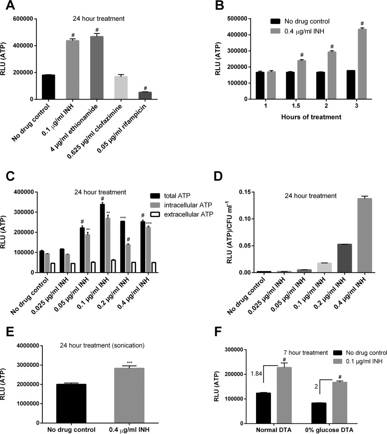
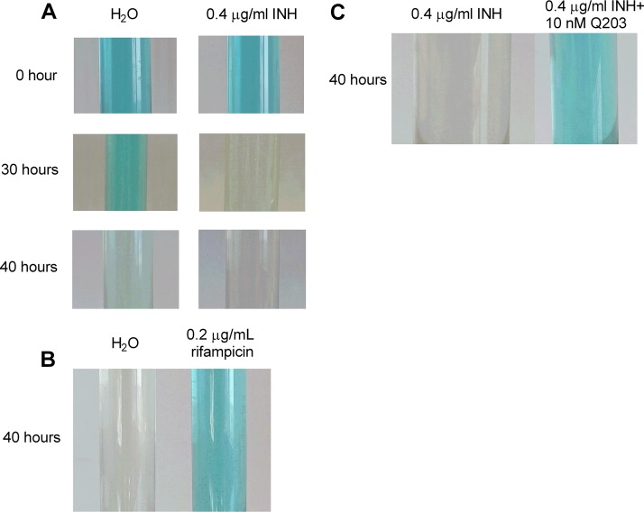
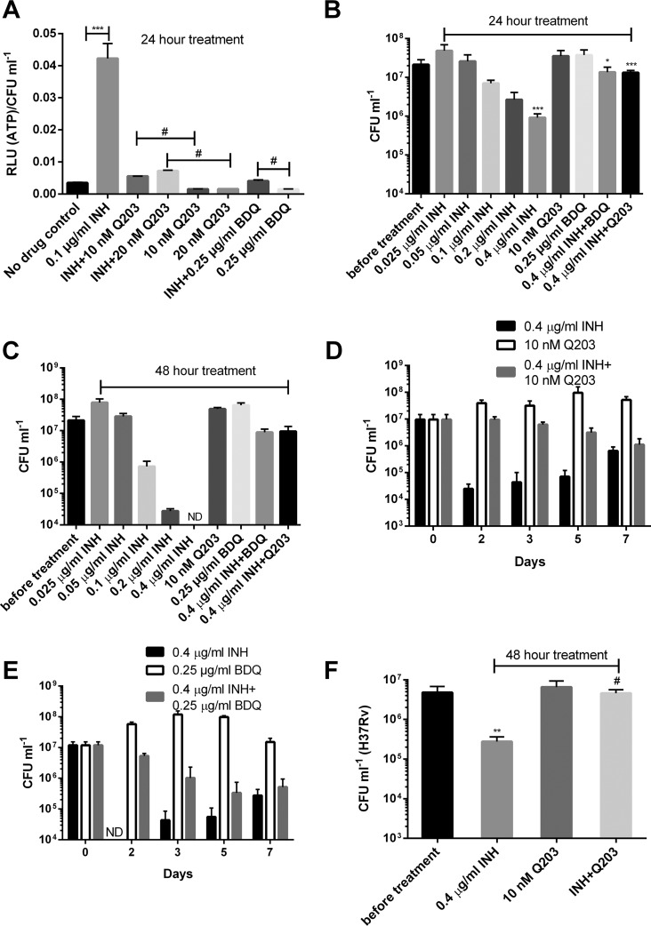
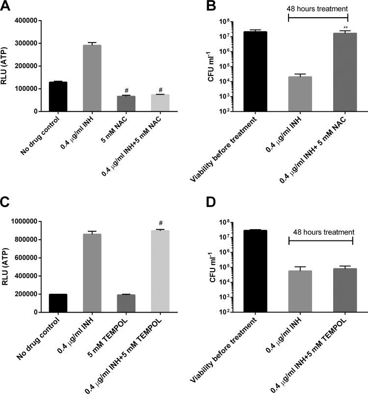
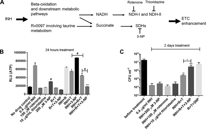
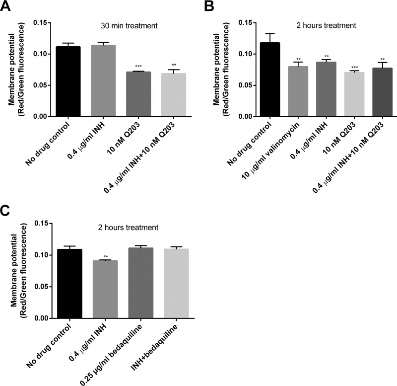
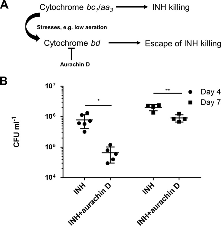
Accumulating evidence suggests that the bactericidal activity of some antibiotics may not be directly initiated by target inhibition. The activity of isoniazid (INH), a key first-line bactericidal antituberculosis drug currently known to inhibit mycolic acid synthesis, becomes extremely poor under stress conditions, such as hypoxia and starvation. This suggests that the target inhibition may not fully explain the bactericidal activity of the drug. Here, we report that INH rapidly increased Mycobacterium bovis BCG cellular ATP levels and enhanced oxygen consumption. The INH-triggered ATP increase and bactericidal activity were strongly compromised by Q203 and bedaquiline, which inhibit mycobacterial cytochrome bc1 and FoF1 ATP synthase, respectively. Moreover, the antioxidant N-acetylcysteine (NAC) but not 4-hydroxy-2,2,6,6-tetramethylpiperidin-1-oxyl (TEMPOL) abrogated the INH-triggered ATP increase and killing. These results reveal a link between the energetic (ATP) perturbation and INH's killing. Furthermore, the INH-induced energetic perturbation and killing were also abrogated by chemical inhibition of NADH dehydrogenases (NDHs) and succinate dehydrogenases (SDHs), linking INH's bactericidal activity further to the electron transport chain (ETC) perturbation. This notion was also supported by the observation that INH dissipated mycobacterial membrane potential. Importantly, inhibition of cytochrome bd oxidase significantly reduced cell recovery during INH challenge in a culture settling model, suggesting that the respiratory reprogramming to the cytochrome bd oxidase contributes to the escape of INH killing. This study implicates mycobacterial ETC perturbation through NDHs, SDHs, cytochrome bc1, and FoF1 ATP synthase in INH's bactericidal activity and pinpoints the participation of the cytochrome bd oxidase in protection against this drug under stress conditions.

Keywords: Mycobacterium tuberculosis; Q203; bedaquiline; electron transport chain; isoniazid; persistence.


Figures

FIG 1
FIG 1
INH and ethionamide enhance cellular ATP. (A) M. bovis BCG cultures in DTA medium were treated with the MICs of various drugs for 24 h before ATP determination. #, P < 0.0001 relative to the no-drug control. (B) ATP kinetics after INH treatment at indicated time points. #, P < 0.0001 relative to the corresponding no-drug control. (C) Bacterial cultures were treated with increasing concentrations of INH for 24 h before measuring extracellular (in the culture filtrate) and intracellular ATP. **, ***, and #, P < 0.01, P < 0.001, and P < 0.0001, respectively, relative to the no-drug control. (D) ATP was determined after INH exposure for 24 h and normalized by viability. (E) Bacterial ATP was determined after sonication. ***, P < 0.001. (F) Cells were grown in DTA medium (with or without glucose) and treated with INH for 7 h prior to ATP measurement. Numbers indicate fold increase of ATP. #, P < 0.0001 relative to the no-drug control. These experiments were performed 2 or 3 times (each in triplicate). Representative data are shown. The error bars indicate standard deviations. RLU, relative light units.
FIG 2
FIG 2
INH enhances oxygen consumption. (A and B) M. bovis BCG cultures were treated with 0.4 µg/ml INH (A) or 0.2 µg/ml rifampin (B), and oxygen consumption was indicated by decolorization of methylene blue (3 µg/ml). (C) Bacterial cultures were treated with 0.4 µg/ml INH (with or without 10 nM Q203), and the oxygen consumption was indicated by methylene blue. Experiments were performed at least 3 times. Representative pictures are shown.
FIG 3
FIG 3
Q203 and bedaquiline abrogate the INH-triggered ATP increase and killing. (A) Normalized ATP levels for M. bovis BCG treated with INH, Q203, or bedaquiline (BDQ). *** and #, P < 0.001 and P < 0.0001, respectively. (B and C) Viability of bacteria determined after 1 (B) and 2 (C) days of treatment with various drugs. *, P < 0.05 relative to the INH group; ***, P < 0.001, relative to viability before treatment for the INH group or to the INH group for the drug combination. ND, not detectable under our plating strategy (<104 CFU/ml). (D and E) Drug kill kinetics following treatment with 0.4 µg/ml INH combined with 10 nM Q203 (D) or 0.25 µg/ml bedaquiline (BDQ) (E). ND, not detectable (<104 CFU/ml). (F) M. tuberculosis H37Rv was treated with 0.4 µg/ml INH and/or 10 nM Q203 for 2 days and viability determined. **, P < 0.01 relative to viability before treatment; #, P < 0.0001 relative to INH group. These experiments were performed 2 or 3 times (each in triplicate). Representative data are shown. The error bars indicate standard deviations.
FIG 4
FIG 4
NAC but not TEMPOL abolishes the INH-induced ATP increase and killing. (A) M. bovis BCG was treated with 0.4 µg/ml INH (with or without 5 mM NAC) for 24 h prior to ATP measurement. #, P < 0.0001 relative to the no-drug control. (B) Viability was determined after exposure to INH (with or without NAC) for 2 days. **, P < 0.01 relative to the INH group. (C) BCG was treated with 0.4 µg/ml INH (with or without 5 mM TEMPOL) for 24 h prior to ATP measurement. #, P < 0.0001 relative to the no-drug control. (D) Viability was determined after exposure to INH (with or without TEMPOL) for 2 days. These experiments were performed 3 times (each in triplicate). Representative data are shown. The error bars indicate standard deviations.
FIG 5
FIG 5
NDH and SDH inhibitors compromise the INH-induced ATP increase and killing. (A) Proposed pathways leading to INH-induced ETC perturbation. Inhibitors targeting the ETC-initiating dehydrogenases are also indicated. (B) M. bovis BCG was treated with various drugs for 24 h before ATP determination. R and T, rotenone and thioridazine, respectively. The concentrations of drugs were used as indicated. *, **, and ***, P < 0.05, P < 0.01, and P < 0.001, respectively, relative to the INH group; #, P < 0.0001 relative to the no-drug control. (C) BCG was challenged with 0.4 µg/ml INH (with or without 200 µM 3-NP, 100 µM rotenone, and 10 µg/ml thioridazine) for 2 days and viability determined. *, P < 0.05; ** and ***, P < 0.001 and P < 0.001, respectively (relative to the INH group). These experiments were performed 3 times (each in triplicate). Representative data are shown. The error bars indicate standard deviations.
FIG 6
FIG 6
INH dissipates mycobacterial membrane potential in an FoF1 ATP synthase-dependent manner. (A and B) M. bovis BCG was treated with INH and/or Q203 for 30 min (A) and 2 h (B) before measuring membrane potential using DiOC2(3). ** and ***, P < 0.01 and P < 0.001, respectively, relative to the no-drug control. The membrane potential was indicated by the ratio of red to green fluorescence. (C) Membrane potential determined in BCG treated with INH and/or bedaquiline for 2 h. **, P < 0.01 relative to the no-drug control. These experiments were performed 3 times (each in triplicate). Representative data are shown. The error bars indicate standard deviations.
FIG 7
FIG 7
Cytochrome bd oxidase inhibition reduces cell recovery in a culture settling model. (A) Dependence of INH’s bactericidal activity on the cytochrome bc1/aa3 supercomplex and escape of INH killing through the activation of cytochrome bd oxidase under stresses such as low aeration. (B) M. bovis BCG (107 CFU/ml) was treated with 0.4 µg/ml INH (with or without 18 µg/ml aurachin D). The cultures were maintained without agitation to allow for cell settling until viability determination at the indicated time points. *and **, P < 0.05 and P < 0.01, respectively. These experiments were performed 3 times (each in triplicate). Results from one representative experiment are shown. The error bars indicate standard deviations.

References

    1. WHO. 2017. Global tuberculosis report 2017. WHO, Geneva, Switzerland.
    1. Zumla A, Nahid P, Cole ST. 2013. Advances in the development of new tuberculosis drugs and treatment regimens. Nat Rev Drug Discov 12:388–404. doi:10.1038/nrd4001. - DOI - PubMed
    1. Al-Saeedi M, Al-Hajoj S. 2017. Diversity and evolution of drug resistance mechanisms in Mycobacterium tuberculosis. Infect Drug Resist 10:333–342. doi:10.2147/IDR.S144446. - DOI - PMC - PubMed
    1. Rawat R, Whitty A, Tonge PJ. 2003. The isoniazid-NAD adduct is a slow, tight-binding inhibitor of InhA, the Mycobacterium tuberculosis enoyl reductase: adduct affinity and drug resistance. Proc Natl Acad Sci U S A 100:13881–13886. doi:10.1073/pnas.2235848100. - DOI - PMC - PubMed
    1. Takayama K, Wang L, David HL. 1972. Effect of isoniazid on the in vivo mycolic acid synthesis, cell growth, and viability of Mycobacterium tuberculosis. Antimicrob Agents Chemother 2:29–35. - PMC - PubMed

Publication types

MeSH terms

LinkOut - more resources