. 2019 Aug 22;201(18):e00741-18.
doi: 10.1128/JB.00741-18. Print 2019 Sep 15.

Flagellar Stators Stimulate c-di-GMP Production by Pseudomonas aeruginosa

Affiliations

Flagellar Stators Stimulate c-di-GMP Production by Pseudomonas aeruginosa

Amy E Baker et al. J Bacteriol. .

Abstract

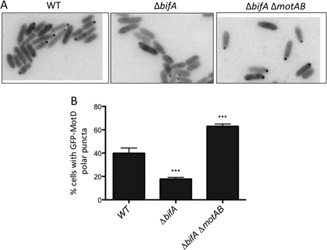
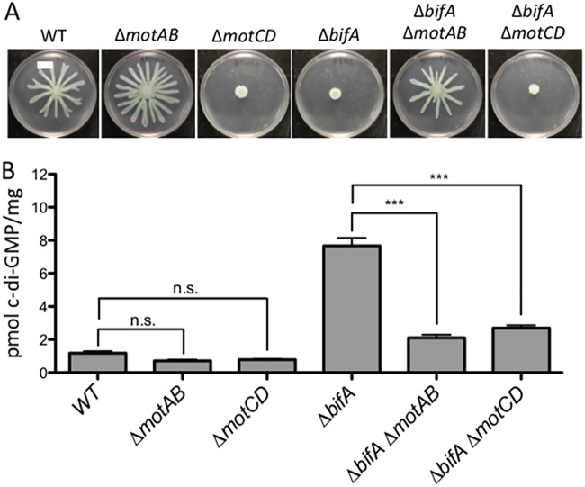
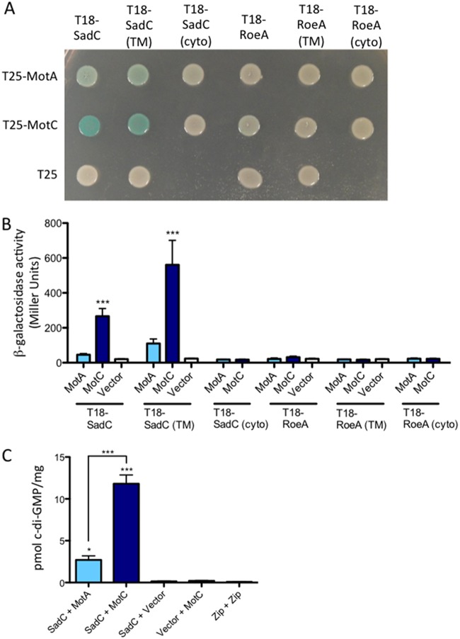
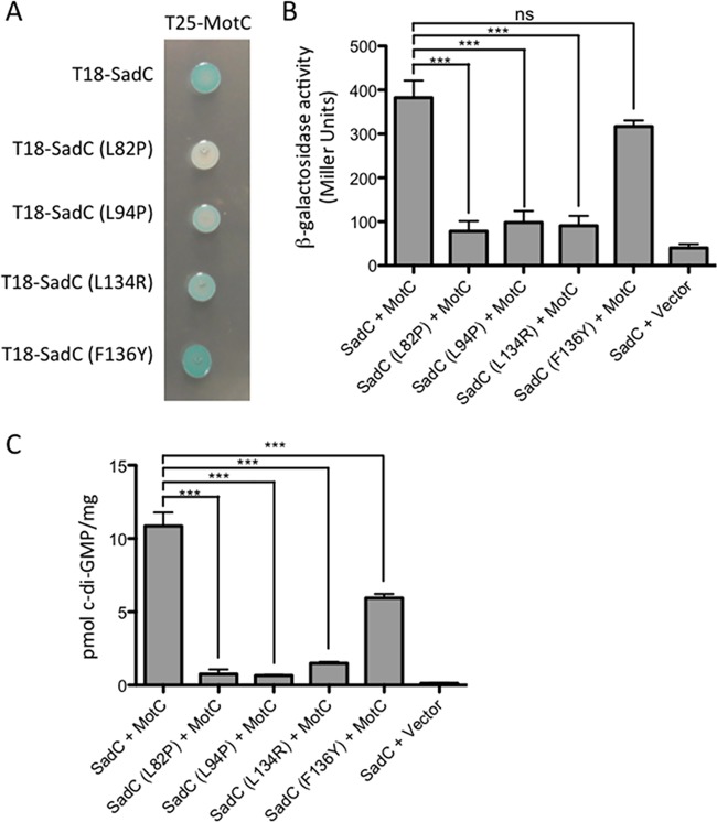
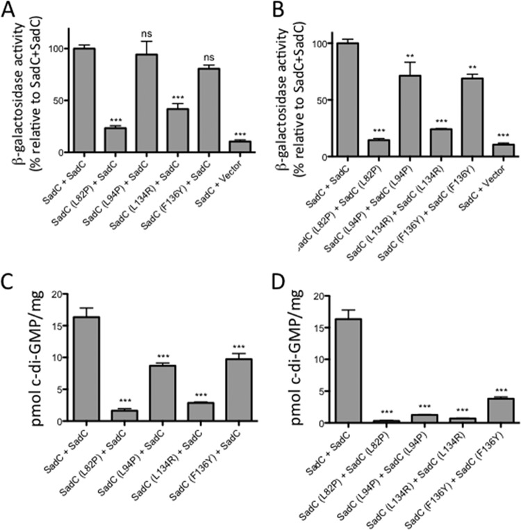
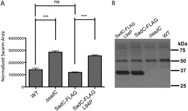
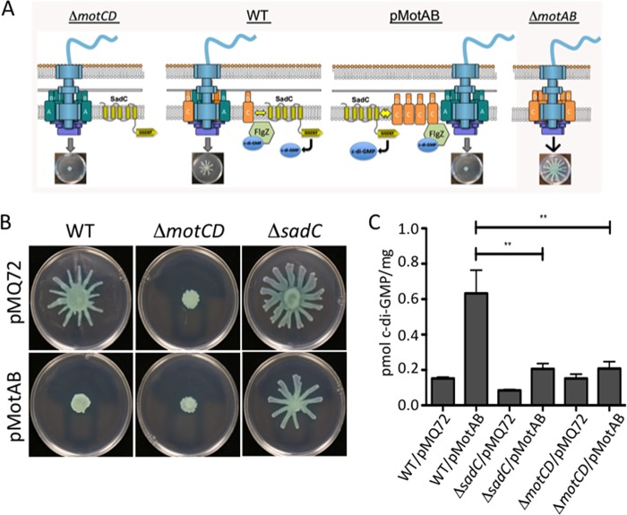
Flagellar motility is critical for surface attachment and biofilm formation in many bacteria. A key regulator of flagellar motility in Pseudomonas aeruginosa and other microbes is cyclic diguanylate (c-di-GMP). High levels of this second messenger repress motility and stimulate biofilm formation. c-di-GMP levels regulate motility in P. aeruginosa in part by influencing the localization of its two flagellar stator sets, MotAB and MotCD. Here, we show that while c-di-GMP can influence stator localization, stators can in turn impact c-di-GMP levels. We demonstrate that the swarming motility-driving stator MotC physically interacts with the transmembrane region of the diguanylate cyclase SadC. Furthermore, we demonstrate that this interaction is capable of stimulating SadC activity. We propose a model by which the MotCD stator set interacts with SadC to stimulate c-di-GMP production under conditions not permissive to motility. This regulation implies a positive-feedback loop in which c-di-GMP signaling events cause MotCD stators to disengage from the motor; then disengaged stators stimulate c-di-GMP production to reinforce a biofilm mode of growth. Our studies help to define the bidirectional interactions between c-di-GMP and the flagellar machinery.IMPORTANCE The ability of bacterial cells to control motility during early steps in biofilm formation is critical for the transition to a nonmotile, biofilm lifestyle. Recent studies have clearly demonstrated the ability of c-di-GMP to control motility via a number of mechanisms, including through controlling transcription of motility-related genes and modulating motor function. Here, we provide evidence that motor components can in turn impact c-di-GMP levels. We propose that communication between motor components and the c-di-GMP synthesis machinery allows the cell to have a robust and sensitive switching mechanism to control motility during early events in biofilm formation.

Keywords: Pseudomonas aeruginosa; biofilm; c-di-GMP; flagella; motility; stator.


Figures

FIG 1
FIG 1
Localization of GFP-MotD is impacted by MotAB. (A) Representative fluorescence microscopy images of the indicated strains expressing GFP-MotD from the chromosome. Grayscale images were inverted using ImageJ. (B) Percentages of cells with GFP-MotD polar puncta for the indicated strains expressing GFP-MotD. These data are from two independent experiments, with at least 200 total cells counted per strain per experiment. Values are reported as means ± standard errors of the means (SEM). Significance was determined by analysis of variance and Dunnett’s posttest for comparison for differences relative to the WT. ***, P < 0.001.
FIG 2
FIG 2
Loss of stators impacts cellular c-di-GMP levels. (A) Representative swarm plates of the strains indicated. (B) Quantification of cellular c-di-GMP levels by LC-MS for the indicated strains grown on swarm plates. Data are expressed as picomoles of c-di-GMP per milligram (dry weight) of cells from which nucleotides were extracted. The data represent results from three independent experiments, each with three biological replicates. Values are reported as means ± SEM. Significance was determined by analysis of variance and Tukey’s post hoc test comparison for differences between strains indicated. n.s., not significant; ***, P < 0.001. As previously reported, the ΔbifA mutant is significantly different from the WT (P < 0.001).
FIG 3
FIG 3
Detection of a physical interaction between MotC and SadC by bacterial two-hybrid analysis. (A) The sadC and roeA genes (and truncated versions) were cloned into the vector pUT18C. “TM” indicates the predicted transmembrane domain, and “cyto” indicates the predicted cytoplasmic region. The motA and motC genes were cloned into the vector pKT25. Plasmids were cotransformed into E. coli BTH101 cells. The transformants were spotted (2 μl) onto LB agar containing Cb, Kan, IPTG, and X-Gal. Plates were incubated at 30°C for 40 h. Cleavage of X-Gal (blue) indicates a positive protein-protein interaction. (B) Bacterial two-hybrid interactions were quantified by measuring β-galactosidase activity of transformants grown at 30°C overnight in LB broth supplemented with Cb and Kan. “Vector” indicates an empty pKT25 vector. The data represent results from 2 experiments, each with 2 to 5 replicates. Values are means ± SEM. Significance was determined by analysis of variance and Dunnett’s posttest comparison for differences from the negative control (T18-SadC’s Vector bar). ***, P < 0.001. (C) Quantification of cellular c-di-GMP levels by LC-MS from B2H assays. The x axis displays the two fusion proteins (listed, in order, as pUT18C and pKT25) cotransformed into BTH101 cells. After being cotransformed with 2 fusion plasmids, cells were spotted onto LB agar containing Cb, Kan, and IPTG (without X-Gal). Plates were incubated at 30°C for 40 h, and then cells were scraped off plates and nucleotides were extracted. Data are expressed as picomoles of c-di-GMP per milligram (dry weight) of cells from which nucleotides were extracted. The data represent results from 2 experiments, each performed in triplicate. Values are means ± SEM. Significance was determined by analysis of variance and Tukey’s post hoc test comparison for differences from the negative control (T18-SadC + Vector) and differences between the indicated samples. Zip + Zip, positive control for the B2H assay. *, P < 0.05; ***, P < 0.001.
FIG 4
FIG 4
Point mutations in the transmembrane domain of SadC disrupt both its interaction with MotC and c-di-GMP production. (A) The wild-type sadC gene and point mutant variants were cloned into the vector pUT18C. The motC gene was cloned into the vector pKT25. Plasmids were cotransformed into E. coli BTH101 cells. The transformants were spotted (2 μl) onto LB agar containing Cb, Kan, IPTG, and X-Gal. Plates were incubated at 30°C for 30 h. Cleavage of X-Gal (blue) indicates a positive protein-protein interaction. (B) Bacterial two-hybrid interactions were quantified by measuring the β-galactosidase activity of transformants grown at 30°C overnight in LB broth supplemented with Cb and Kan. “Vector” indicates an empty pKT25 vector. The data represent results from two experiments, each performed in triplicate. Values are means ± SEM. Significance was determined by analysis of variance and Dunnett’s posttest comparison for difference from the wild-type interaction (T18-SadC plus T25-MotC). ***, P < 0.001; ns, not significant. (C) Quantification of cellular c-di-GMP levels by LC-MS from B2H assays. The x axis displays the two fusion proteins (listed, in order, as pUT18C and pKT25) cotransformed into BTH101 cells. After being cotransformed with 2 fusion plasmids, cells were spotted onto LB agar containing Cb, Kan, and IPTG (with no X-Gal). Plates were incubated at 30°C for 40 h, and then cells were scraped off plates and nucleotides were extracted. Data are expressed as picomoles of c-di-GMP per milligram (dry weight) of cells from which nucleotides were extracted. Data represent results from three experiments, each performed in triplicate. Values are means ± SEM. Significance was determined by analysis of variance and Tukey’s post hoc test comparison for differences between the strains indicated. ***, P < 0.001.
FIG 5
FIG 5
TM domain mutations in SadC impact dimerization and c-di-GMP production. (A, B) The wild-type sadC gene was cloned into the vectors pUT18C and pKT25, and mutant sadC plasmids were generated using in vitro site-directed mutagenesis. Plasmids were cotransformed into E. coli BTH101 cells. The transformants were spotted (2 μl) onto LB agar containing Cb, Kan, and IPTG. Plates were incubated at 30°C for 30 h. Bacterial two-hybrid interactions were quantified by measuring β-galactosidase activity in transformants grown at 30°C overnight in LB broth supplemented with Cb and Kan. “Vector” indicates an empty pKT25 vector. Calculated Miller units are represented as percentages of the SadC-SadC interaction (set at 100%). Data are from 2 experiments, each performed in triplicate. Values are means ± SEM. Significance was determined by analysis of variance and Dunnett’s posttest comparison for differences from the SadC-SadC interaction. **, P < 0.01; ***, P < 0.001; ns, not significant. (C, D) Quantification of cellular c-di-GMP levels by LC-MS from B2H assays. The x axis displays the two fusion proteins (listed, in order, as pUT18C and pKT25) cotransformed into BTH101 cells. After being cotransformed with 2 fusion plasmids, cells were spotted onto LB agar containing Cb, Kan, and IPTG (with no X-Gal). Plates were incubated at 30°C for 40 h, and then cells were scraped off plates and nucleotides were extracted. Data are expressed as picomoles of c-di-GMP per milligram (dry weight) of cells from which nucleotides were extracted. Data in panels C and D are from three experiments, each performed in triplicate. Values are means ± SEM. Significance was determined by analysis of variance and Dunnett’s posttest comparison for differences from the wild-type interaction (T18-SadC plus T25-SadC). ***, P < 0.001.
FIG 6
FIG 6
Analysis of SadC point mutations in P. aeruginosa. (A) Swarming motility as assessed for the indicated strains. Significance was determined by analysis of variance and Tukey’s post hoc test comparison for differences from the WT or the SadC-FLAG expression strain swarm zone, as indicated. ***, P < 0.0001; ns, not significant. (B) Western blot analysis of the WT and mutant FLAG-tagged SadC. The positions of the molecular weight size standards are shown on the right. The bands at ∼50 and ∼80 kDa are nonspecific, cross-reacting bands and serve as additional loading controls. Each lane contains 250 μg of crude cell extract of the P. aeruginosa carrying the indicated allele of the SadC or SadC-FLAG gene.
FIG 7
FIG 7
MotAB-MotCD dynamics impact c-di-GMP levels. (A) Shown is a model of the flagellar motor with various configurations of the stators, including a ΔmotCD mutant (left), the wild type (center, left), a strain expressing MotAB from a multicopy plasmid (center, right), and the ΔmotAB mutant (left). Also shown is the c-di-GMP effector FlgZ, which can interact with MotC when FlgZ is in the c-di-GMP-bound state. A, B, C, and D refer to MotA, MotB, MotC and MotD, respectively. (B) Swarming motility as assessed for the indicated strains. (C) c-di-GMP quantification assays. Data are expressed as picomoles of c-di-GMP per milligram (dry weight) of cells from which nucleotides were extracted. Values are means ± SEM. Significance was determined by analysis of variance and Dunnett’s posttest comparison to the WT carrying the vector plasmid (pMQ72). **, P < 0.01.

References

    1. Hengge R. 2009. Principles of c-di-GMP signalling in bacteria. Nat Rev Microbiol 7:263–273. doi:10.1038/nrmicro2109. - DOI - PubMed
    1. Romling U, Galperin MY, Gomelsky M. 2013. Cyclic di-GMP: the first 25 years of a universal bacterial second messenger. Microbiol Mol Biol Rev 77:1–52. doi:10.1128/MMBR.00043-12. - DOI - PMC - PubMed
    1. Merritt JH, Brothers KM, Kuchma SL, O'Toole GA. 2007. SadC reciprocally influences biofilm formation and swarming motility via modulation of exopolysaccharide production and flagellar function. J Bacteriol 189:8154–8164. doi:10.1128/JB.00585-07. - DOI - PMC - PubMed
    1. Merritt JH, Ha DG, Cowles KN, Lu W, Morales DK, Rabinowitz J, Gitai Z, O'Toole GA. 2010. Specific control of Pseudomonas aeruginosa surface-associated behaviors by two c-di-GMP diguanylate cyclases. mBio 1:e00183-10. doi:10.1128/mBio.00183-10. - DOI - PMC - PubMed
    1. Doyle TB, Hawkins AC, McCarter LL. 2004. The complex flagellar torque generator of Pseudomonas aeruginosa. J Bacteriol 186:6341–6350. doi:10.1128/JB.186.19.6341-6350.2004. - DOI - PMC - PubMed

Publication types

MeSH terms

LinkOut - more resources