. 2019 Jan 14;5(1):1.
doi: 10.1038/s41537-018-0069-1.

Region-specific inhibition of 14-3-3 proteins induces psychomotor behaviors in mice

Affiliations

Region-specific inhibition of 14-3-3 proteins induces psychomotor behaviors in mice

Kourtney Graham et al. NPJ Schizophr. .

Abstract

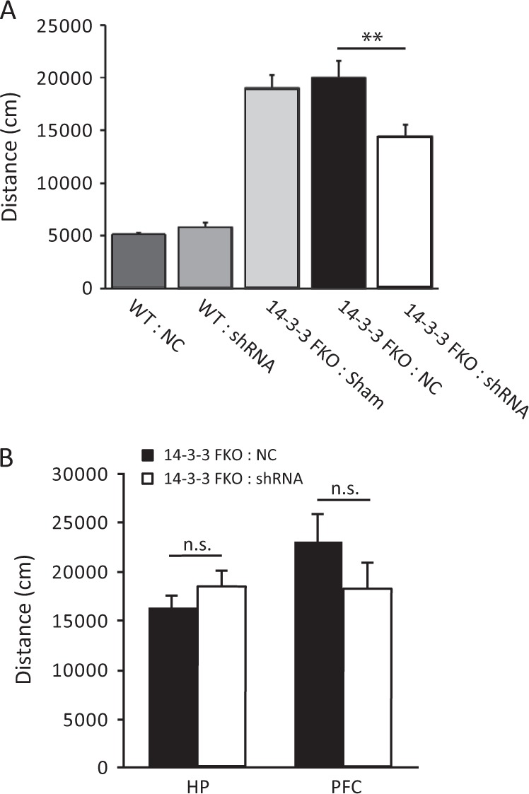
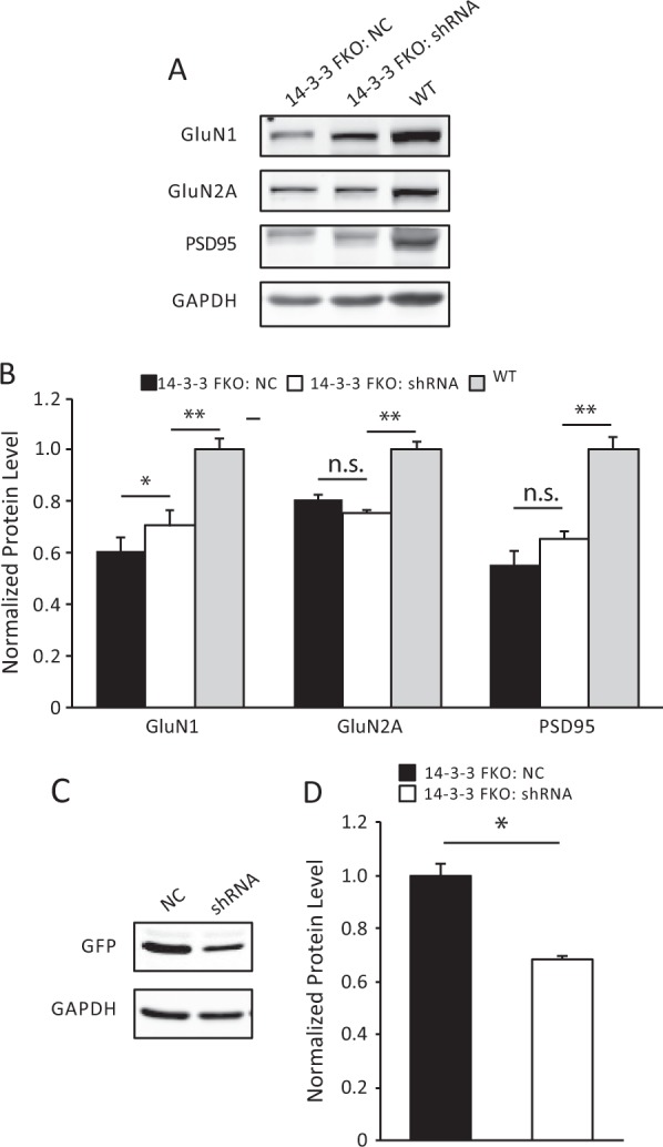
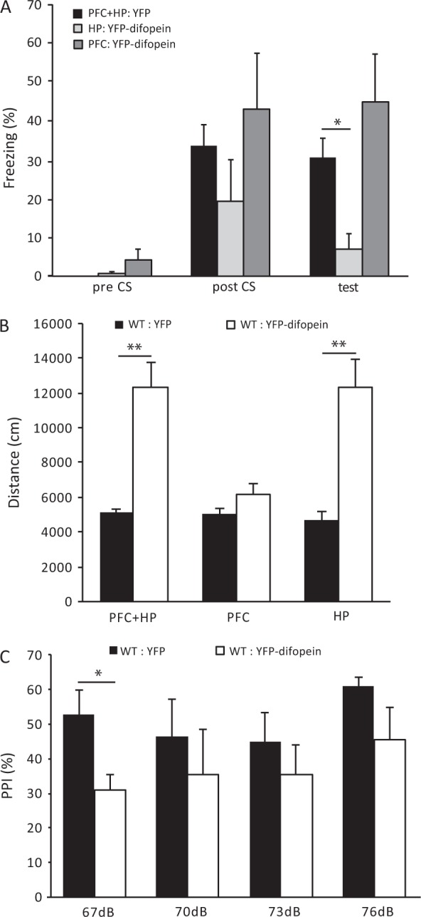
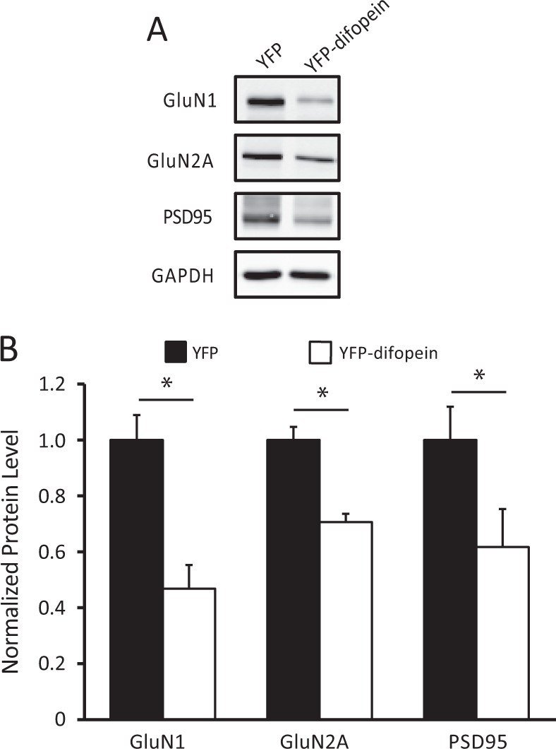
The 14-3-3 family of proteins is genetically linked to several psychiatric disorders, including schizophrenia. Our 14-3-3 functional knockout (FKO) mice, as well as other 14-3-3 knockout models, have been shown to exhibit behavioral endophenotypes related to schizophrenia. While specific forebrain regions, such as the prefrontal cortex (PFC) and hippocampus (HP), have been implicated in schizophrenic pathophysiology, the role of these brain regions in the top-down control of specific schizophrenia-associated behaviors has not been examined. Here, we used an adeno-associated virus (AAV) delivered shRNA to knock down the expression of the 14-3-3-inhibitor transgene, thus selectively restoring the function of 14-3-3 in the forebrain of the 14-3-3 FKO mice, we found that injection of the AAV-shRNA into both the PFC and the HP is necessary to attenuate psychomotor activity of the 14-3-3 FKO mice. Furthermore, we found that acute inhibition of 14-3-3, through the delivery of an AAV expressing the 14-3-3 inhibitor to both the PFC and HP, can trigger psychomotor agitation. Interestingly, when assessing the two brain regions separately, we determined that AAV-mediated expression of the 14-3-3 inhibitor specifically within the HP alone is sufficient to induce several behavioral deficits including hyperactivity, impaired associative learning and memory, and reduced sensorimotor gating. In addition, we show that post-synaptic NMDA receptor levels are regulated by acute 14-3-3 manipulations. Taken together, findings from this study directly link 14-3-3 inhibition in specific forebrain regions to certain schizophrenia-associated endophenotypes.


Conflict of interest statement

The authors declare no competing interests.

Figures

Fig. 1
Fig. 1
Region-specific knockdown of the transgene expression in 14-3-3 FKO mice. a Representative image of YFP (green) expression in tsA 201 cells transfected with YFP-difopein cDNA. b Representative images of reduced YFP expression in cells co-transfected with cDNAs of YFP-difopein and pSicoR-mCherry-shRNA. c Schematic map of recombinant adeno-associated viral (rAAV) vector used to co-express the shRNA against YFP (shRNA) and mCherry proteins. d Representative image of a sagittal section of a 14-3-3 FKO mouse brain injected with AAV-mCherry-shRNA (red) in both the prefrontal cortex (PFC) e and dorsal hippocampus (HP) f. Virus (red) and difopein transgene (green) expression were detected using fluorescent and confocal microscopy. There is minimal colocalization between red and green signals in the PFC (EI−III) or HP CA1 (FI−III), indicating a knockdown of difopein expression in AAV-infected cells. g, h Representative images of the PFC (GI−III) and the HP CA1 (HI−III) of the 14-3-3 FKO mice injected with the negative control (NC) AAV expressing a scrambled shRNA and mCherry. There is colocalization of red and green signals in both the PFC and HP regions. Scale bars represent 300 μm d and 100 μm eh
Fig. 2
Fig. 2
Attenuation of hyperlocomotive behavior in the 14-3-3 FKO mice. a Bilateral injection of the AVV-mCherry-YFP/shRNA to both the PFC and HP of 14-3-3 FKO mice (14-3-3 FKO: shRNA) (n = 12) significantly decreases distance traveled during open field testing, compared with 14-3-3 FKO mice injected with control virus injections in the same brain regions (14-3-3 FKO: NC) (n = 15; F(1, 25) = 12.57; p = 0.002). Distance traveled is similar in both WT mice with control virus injections (n = 6) and WT mice with shRNA injections (n = 10; F(1, 14) = 1.892; p = 0.191). b Bilateral injections of AAV-mCherry-YFP/shRNA to either the HP alone (n = 7; F(1, 10) = 0.836; p = 0.382) or PFC alone (n = 6; F(1, 10) = 1.516; p = 0.246) were not able to attenuate the locomotor activity in open field test when compared with 14-3-3 FKO mice injected with scrambled shRNA control (HP, n = 5; PFC, n = 6). Data are presented as mean ± S.E.M, with statistical significance denoted as: n.s., not significant; **p < 0.01; one-way ANOVA
Fig. 3
Fig. 3
Elevated levels of post-synaptic density proteins induced by region-specific knockdown of the 14-3-3 inhibitor. a, c Representative images of western blots from dorsal hippocampal PSD fractions a and cell lysate c and after bilateral injections of AAV-mCherry-YFP/shRNA (shRNA) or the negative control AAV (NC) in the 14-3-3 FKO mice. b When compared with NC injections (n = 6), there is a significant increase in the GluN1 (F(1, 10) = 6.74, p = 0.048) subunit as well as an upward trend in PSD95 (F(1, 10) = 4.32, p = 0.09) in the PSD after bilateral injections of shRNA to the 14-3-3 FKO mice (n = 6). The levels of GluN1, GluN2A, and PSD95 in the shRNA group are lower than that in WT group (GluN1: F(1, 10) = 21.78, p = 0.006; GluN2A: F(1, 10) = 63.79, p < 0.001; PSD95: F(1, 10) = 29.87, p = 0.003). d AAV-mCherry-YFP/shRNA injections (n = 6) significantly reduced the levels of YFP-difopein in the dorsal hippocampus of 14-3-3 FKO mice, compared with controls (n = 6) (F(1, 10) = 14.54, p = 0.013). Blots were probed with GAPDH as a loading control. Protein levels were normalized to that of 14-3-3 FKO mice injected with control virus b or that of WT mice d. Data are presented as mean ± S.E.M., with statistical significance denoted as: n.s. = not significant, *p < 0.05, **p < 0.01; two-tailed t-test
Fig. 4
Fig. 4
Region-specific expression of a 14-3-3 inhibitor in WT mice. a Schematic map of the recombinant adeno-associated viral (AAV) vector used to express the YFP-difopein under control of the CamKII promoter. b Representative images of a sagittal mouse brain section with YFP-difopein viral injections in both the PFC (BI) and dorsal HP (BII). YFP-difopein expression (green) was detected by fluorescent and confocal microscopy. Scale bars represent 300 and 100 μm respectively
Fig. 5
Fig. 5
Behavioral deficits induced by YFP-difopein expression in WT mice. a Compared with WT mice injected with AAV-YFP in both the HP and PFC (n = 14), bilateral injections of AAV-YFP-difopein to the HP alone significantly reduce freezing behavior in contextual fear conditioning test 24 h after training (n = 6; F(1, 18) = 6.994; p = 0.016). However, injections of AAV-YFP-difopein to PFC alone does not affect freezing behavior when compared with WT controls (n = 6; F(1, 18) = 1.505; p = 0.236). b Bilateral injections of AAV-YFP-difopein into the HP, either alone (n = 8; F(1, 7.34) = 27.826; p = 0.001) or together with injection to the PFC (n = 9; F(1, 9.668) = 17.64; p = 0.002), significantly increase travel distances in open field test, compared with WT mice injected with AAV-YFP (HP: n = 9, HP + PFC: n = 6). However, injections of AAV-YFP-difopein to the PFC alone (n = 7; F(1, 10) = 1.953; p = 0.192) did not affect locomotive behavior of WT mice. c Bilateral injections of AAV-YFP-difopein to the HP of WT mice causes a decrease in pre-pulse inhibition (PPI) at all pre-pulse levels tested, while PPI is significantly lower at the pre-pulse level of 67 dB (n = 5; F (1, 8) = 6.746; p = 0.032), compared with WT mice with AAV-YFP injected to the HP of (n = 5). Data are presented as mean ± S.E.M. with statistical significance denoted as: *p < 0.05; **p < 0.01; one-way ANOVA
Fig. 6
Fig. 6
Reduction of NMDA receptor subunits in the post-synaptic density induced by YFP-difopein expression. a Representative images of western blots from dorsal hippocampal PSD fractions of WT mice injected with either control virus (AAV-YFP) or AAV-YFP-difopein virus. b Compared with that of controls (n = 6 for all groups), there is a significant decrease in the levels of GluN1 (n = 6, F (1, 10) = 14.60, p = 0.012), GluN2A (n = 6, F(1, 10) = 12.50, p = 0.017) subunits and the levels of PSD95 (n = 6, F(1, 10) = 11.73, p = 0.019) in YFP-difopein injected mice. Blots were probed with GAPDH as a loading control and protein levels were normalized to that of WT control. Data are presented as mean ± S.E.M., with statistical significance *p < 0.05, two-tailed t-test

Similar articles

Cited by

References

    1. Volk DW, Lewis DA. Prefrontal cortical circuits in schizophrenia. Curr. Top. Behav. Neurosci. 2010;4:485–508. doi: 10.1007/7854_2010_44. - DOI - PubMed
    1. Small SA, Schobel SA, Buxton RB, Witter MP, Barnes CA. A pathophysiological framework of hippocampal dysfunction in ageing and disease. Nat. Rev. Neurosci. 2011;12:585–601. doi: 10.1038/nrn3085. - DOI - PMC - PubMed
    1. Ross CA, Margolis RL, Reading SA, Pletnikov M, Coyle JT. Neurobiology of schizophrenia. Neuron. 2006;52:139–153. doi: 10.1016/j.neuron.2006.09.015. - DOI - PubMed
    1. Rasetti R, et al. Altered cortical network dynamics: a potential intermediate phenotype for schizophrenia and association with ZNF804A. Arch. Gen. Psychiatry. 2011;68:1207–1217. doi: 10.1001/archgenpsychiatry.2011.103. - DOI - PubMed
    1. Meyer-Lindenberg AS, et al. Regionally specific disturbance of dorsolateral prefrontal-hippocampal functional connectivity in schizophrenia. Arch. Gen. Psychiatry. 2005;62:379–386. doi: 10.1001/archpsyc.62.4.379. - DOI - PubMed