. 2019 Jan 14;9(1):85.
doi: 10.1038/s41598-018-36347-7.

Behaviors of Glioblastoma Cells in in Vitro Microenvironments

Affiliations

Behaviors of Glioblastoma Cells in in Vitro Microenvironments

Wenwen Diao et al. Sci Rep. .

Abstract

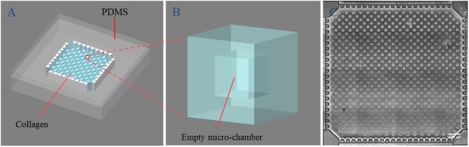
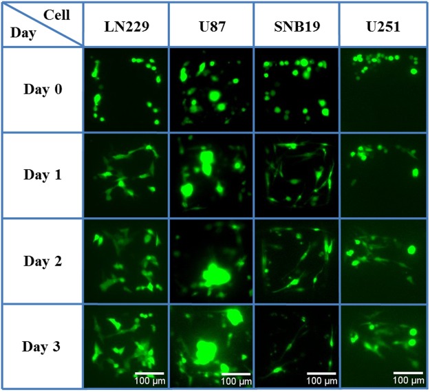
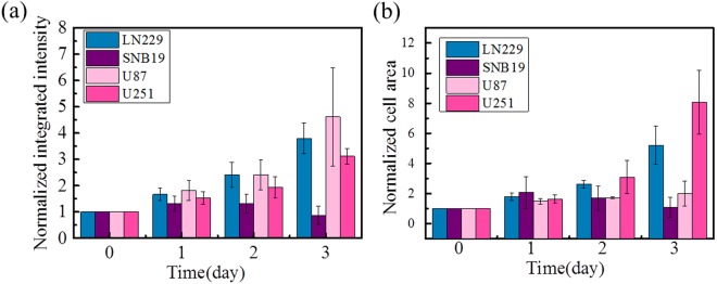
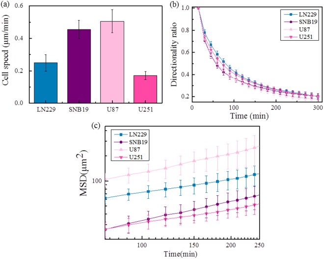
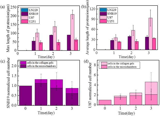
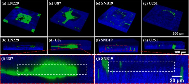
Glioblastoma (GBM) is the most malignant and highly aggressive brain tumor. In this study, four types of typical GBM cell lines (LN229, SNB19, U87, U251) were cultured in a microfabricated 3-D model to study their in vitro behaviors. The 3-D in vitro model provides hollow micro-chamber arrays containing a natural collagen interface and thus allows the GBM cells to grow in the 3-D chambers. The GBM cells in this model showed specific properties on the aspects of cell morphology, proliferation, migration, and invasion, some of which were rarely observed before. Furthermore, how the cells invaded into the surrounding ECM and the corresponding specific invasion patterns were observed in details, implying that the four types of cells have different features during their development in cancer. This complex in vitro model, if applied to patient derived cells, possesses the potential of becoming a clinically relevant predictive model.


Conflict of interest statement

The authors declare no competing interests.

Figures

Figure 1
Figure 1
(A) 3D in vitro model with micro-chambers in collagen; (B) enlarged image showing one micro-chamber; (C) top view of the chip, captured in the bright field and processed with ImageJ, where the scale bar is 1000 μm. The size of the microchambers is 200 × 200 μm2, with the distance between nearest neighbor pairs being 400 μm.
Figure 2
Figure 2
Representative fluorescent images showing the morphology of the four types of GBM cells in micro-chambers within collagen during the four culture days.
Figure 3
Figure 3
Cell proliferation rate of LN229 (blue), SNB19 (purple), U87 (light pink), and U251 (dark pink) indicated by (a) normalized integrated intensity and (b) normalized cell area. Data are presented as mean ± SD of three independent experiments.
Figure 4
Figure 4
Cell migration speed, directionality ratio, and MSD of four types of GBM cells: (a) the average cell migration speed (N = 200 for each test); (b) directionality ratio (N = 20 for each test); (c) mean square displacement (MSD), plotted as function of time interval (N = 20 for each test). Data are presented as mean ± SD of three independent experiments.
Figure 5
Figure 5
(a) Average and (b) maximum protrusion length of four types of GBM cells; (c) normalized cell number (conjectured from normalized integrated fluorescent intensity) of SNB19 cells inside individual micro-chambers and those invading into the surrounding collagen gel for four consecutive days; (d) normalized cell number (integrated intensity) of U87 cells inside individual micro-chambers and those in the surrounding gel. Data are presented as mean ± SD of three independent experiments.
Figure 6
Figure 6
Confocal images showing representative cell filopodia protrusions (green fluorescent) in a single micro-chamber, where the top and bottom rows are, respectively, top-view and side-view images. (i,j) Locally amplified side-view images of (d) and (f). The collagen fibers (blue) were observed in reflection mode.

Similar articles

Cited by

References

    1. Ostrom QT, et al. CBTRUS Statistical Report: Primary Brain and Central Nervous System Tumors Diagnosed in the United States in 2006-2010. Neuro-Oncology. 2013;15:1–56. doi: 10.1093/neuonc/not151. - DOI - PMC - PubMed
    1. Furnari FB, et al. Malignant astrocytic glioma: genetics, biology, and paths to treatment. Gene Dev. 2007;21:2683–2710. doi: 10.1101/gad.1596707. - DOI - PubMed
    1. Ho, I. A. W. & Shim, W. S. N. Contribution of the Microenvironmental Niche to Glioblastoma Heterogeneity. Biomed Res Int, 1155/2017/9634172 (2017). - PMC - PubMed
    1. Xiao W, Sohrabi A, Seidlits SK. Integrating the glioblastoma microenvironment into engineered experimental models. Future science OA. 2017;3:FSO189. doi: 10.4155/fsoa-2016-0094. - DOI - PMC - PubMed
    1. Gieryng A, Pszczolkowska D, Walentynowicz KA, Rajan WD, Kaminska B. Immune microenvironment of gliomas. Lab Invest. 2017;97:498–518. doi: 10.1038/labinvest.2017.19. - DOI - PubMed

Publication types