. 2019 Jan 14;9(1):84.
doi: 10.1038/s41598-018-36452-7.

Identification of extracellular vesicles and characterization of miRNA expression profiles in human blastocoel fluid

Affiliations

Identification of extracellular vesicles and characterization of miRNA expression profiles in human blastocoel fluid

R Battaglia et al. Sci Rep. .

Abstract

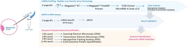
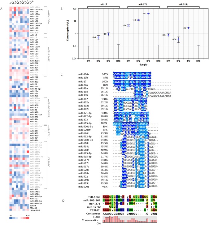
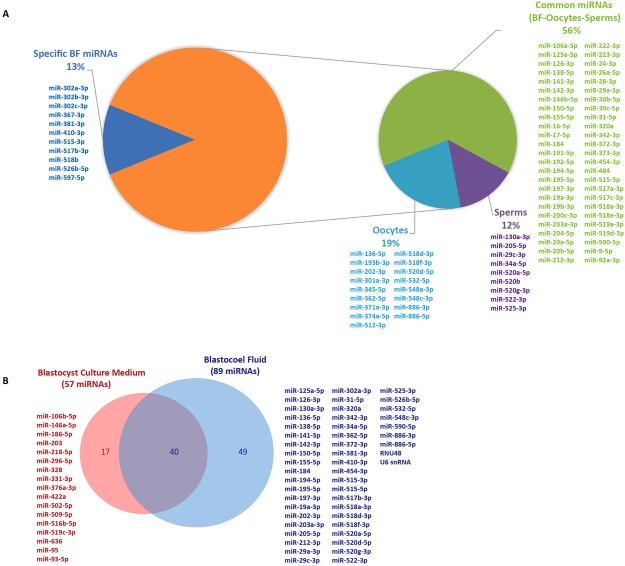
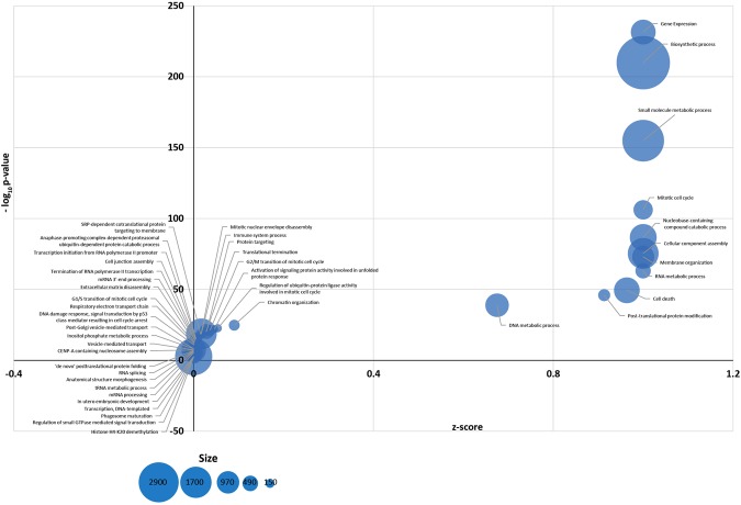
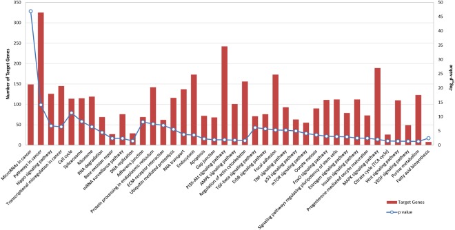
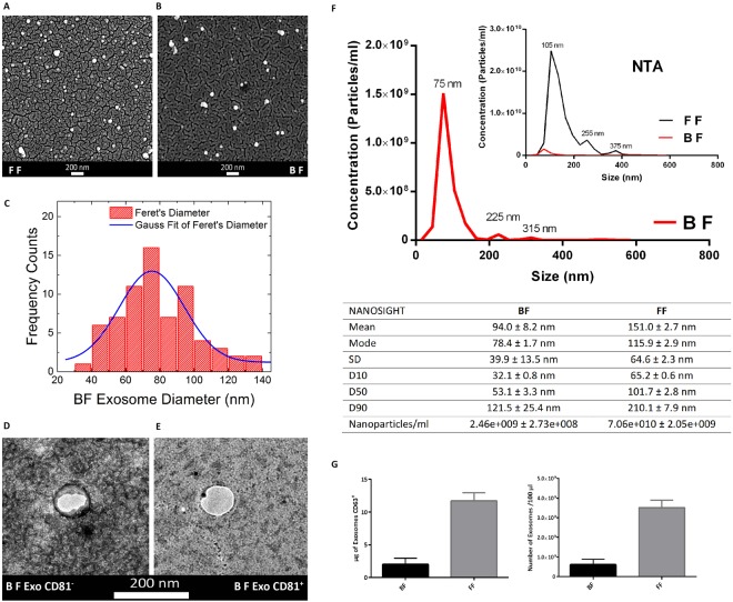
In this study, for the first time, we demonstrated the presence of microRNAs and extracellular vesicles in human blastocoel fluid. The bioinformatic and comparative analyses identified the biological function of blastocoel fluid microRNAs and suggested a potential role inside the human blastocyst. We found 89 microRNAs, expressed at different levels, able to regulate critical signaling pathways controlling embryo development, such as pluripotency, cell reprogramming, epigenetic modifications, intercellular communication, cell adhesion and cell fate. Blastocoel fluid microRNAs reflect the miRNome of embryonic cells and their presence, associated with the discovery of extracellular vesicles, inside blastocoel fluid, strongly suggests their important role in mediating cell communication among blastocyst cells. Their characterization is important to better understand the earliest stages of embryogenesis and the complex circuits regulating pluripotency. Moreover, blastocoel fluid microRNA profiles could be influenced by blastocyst quality, therefore, microRNAs might be used to assess embryo potential in IVF cycles.


Conflict of interest statement

The authors declare no competing interests.

Figures

Figure 1
Figure 1
Schematic overview of experimental workflow for miRNA profiling and exosome characterization in human Blastocoel Fluid.
Figure 2
Figure 2
miRNA expression in human Blastocoel Fluid (BF). (A) Heat map representation of normalized expression data (−ΔCT values) for 89 miRNAs from nine BF samples. The red and blue colors represent miRNA expression levels. (B) Quantification of BF miRNAs using droplet digital PCR (ddPCR). Measurements for miR-17, miR-372 and miR-519d are shown as miRNA copies/μl ddPCR mix. All NTC controls do not show positive droplets. (C) Alignment of human BF miRNAs. Blue shading indicates nucleotides of miRNA sequences with identical positions in the alignment. Light blue shading indicates nucleotides conserved in the major groups of BF miRNAs. (D) Consensus clustering and frequency of nucleotide conservation for groups of BF-miRNAs. Most of the mature miRNAs conserve the seed sequence AAGUGC.
Figure 3
Figure 3
Comparison of BF miRNA expression profile in human oocyte, sperm and blastocyst culture medium. (A) Pie charts representing miRNAs from BF identified in human oocytes and sperms. Most of the detected miRNAs (87%) were shared among human gametes. Their distribution is indicated in the small Pie chart. (B) Venn diagram comparing BF miRNA expression with culture medium miRNAs.
Figure 4
Figure 4
Significant GOs, biological processes, for miRNAs identified in human BFs. Bubble chart representing the most enriched functional categories for miRNA targets in BF. The y-axis represents the -log10 (P-value) and the x-axis the corresponding z-scores. The radius of the bubble is proportional to the size of the mRNA functional category (in terms of number of genes).
Figure 5
Figure 5
Signaling Pathway enrichment analysis for BF miRNAs with KEGG. Histograms representing pathways enriched in BF miRNA target genes. The probability values are reported as −log10 (P-value).
Figure 6
Figure 6
Morphological and Molecular Characterization of Exosomes from human Blastocoel Fluid. (A,B) Scanning Electron Micrographs of extracellular vesicles isolated from Follicular Fluid (FF) and Blastocoel Fluid (BF) respectively. (C) Diameter distribution of exosomes from BFs. Gauss fit of the Feret’s diameter histogram measured on SEM microscopies show an average BF diameter of 75 ± 3 nm and a full width at half maximum (FWHM) of 38 ± 8 nm. (D) Transmission Electron Microscopy images of exosomes from BFs. (E) Transmission Electron Microscopy images of exosomes from BFs marked with CD81. (F) Nanoparticle Tracking Analysis (NTA) of BF extracellular vesicles. Extracellular vesicles from Follicular Fluid were used as reference control (inset). Diameters and concentration of vesicles are indicated in the table. (G) ELISA assay with the tetraspanin CD63 antibody of BF exosomes. Amount (µg) of CD63 protein and EV concentration (number of particles/100 μl) evaluated in BFs. Follicular Fluid (FF) samples were used as reference control. Results are expressed as mean ± SEM.

References

    1. Hammond ER, Shelling AN, Cree LM. Nuclear and mitochondrial DNA in blastocoele fluid and embryo culture medium: evidence and potential clinical use. Hum Reprod. 2016;31:1653–61. doi: 10.1093/humrep/dew132. - DOI - PubMed
    1. Sepúlveda SJ, Portella JR, Noriega LP, Escudero EL, Noriega LH. Extendedculture up to the blastocyst stage: a strategy to avoid multiple pregnancies inassisted reproductive technologies. Biol Res. 2011;44:195–9. doi: 10.4067/S0716-97602011000200012. - DOI - PubMed
    1. Richardson A, et al. A clinically useful simplified blastocyst grading system. Reprod Biomed Online. 2015;31:523–30. doi: 10.1016/j.rbmo.2015.06.017. - DOI - PubMed
    1. Gardner DK, Schoolcraft WB. Culture and transfer of human blastocysts. Curr Opin Obstet Gynecol. 1999;11:307–11. doi: 10.1097/00001703-199906000-00013. - DOI - PubMed
    1. Veeck LL, et al. High pregnancy rates can be achieved after freezing and thawinghuman blastocysts. Fertil Steril. 2004;82:1418–27. doi: 10.1016/j.fertnstert.2004.03.068. - DOI - PubMed

Publication types