. 2019 May;38(18):3415-3427.
doi: 10.1038/s41388-018-0652-y. Epub 2019 Jan 14.

Cancer therapeutic targeting using mutant-p53-specific siRNAs

Affiliations

Cancer therapeutic targeting using mutant-p53-specific siRNAs

Ifeoma Ubby et al. Oncogene. 2019 May.

Abstract

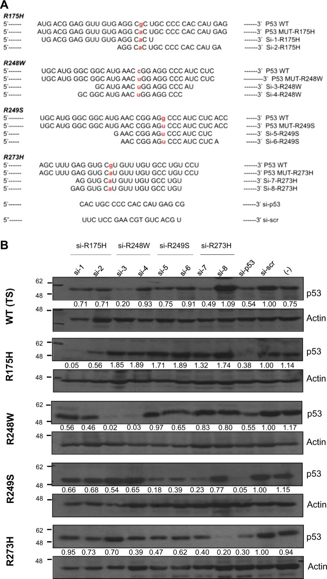
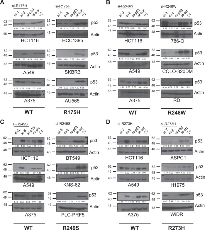
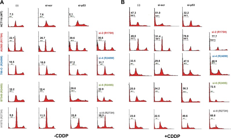
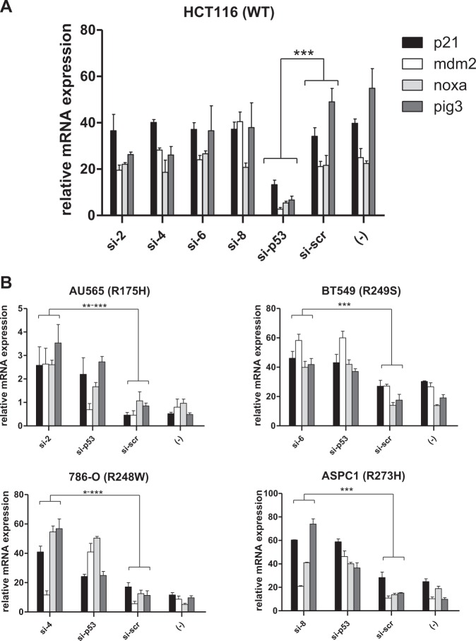
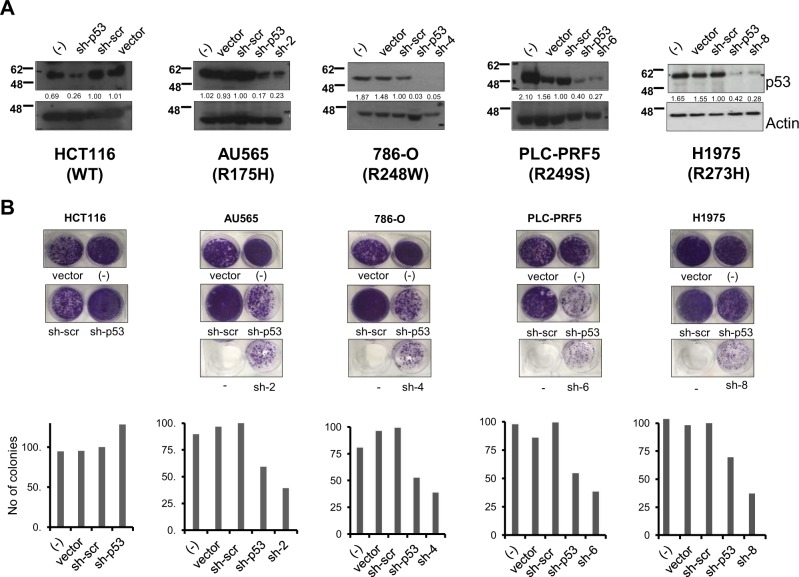
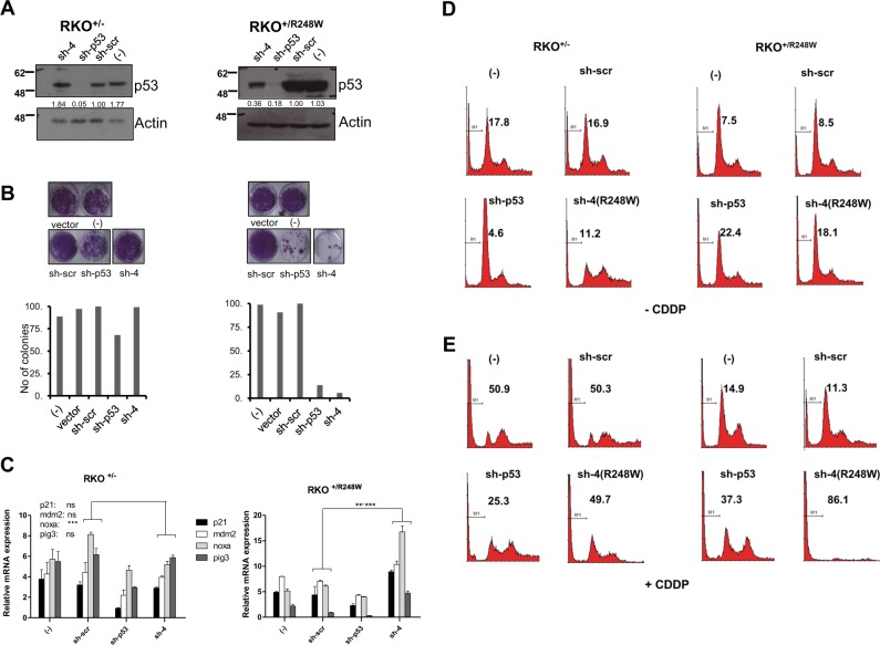
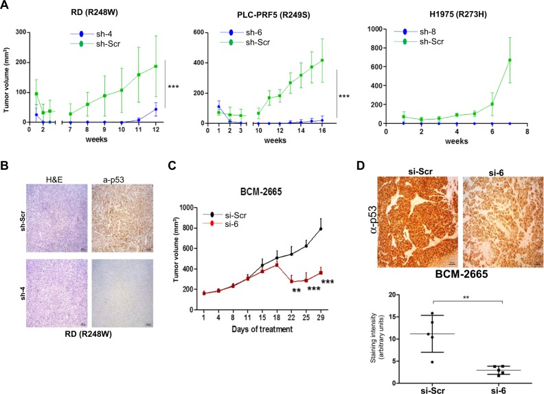
Mutations in Tp53 compromise therapeutic response, due either to the dominant-negative effect over the functional wild-type allele; or as a result of the survival advantage conferred by mutant p53 to which cancer cells become addicted. Thus, targeting mutant p53 represents an effective therapeutic strategy to treat over half of all cancers. We have therefore generated a series of small-interfering-RNAs, capable of targeting four p53 hot-spot mutants which represent ~20% of all p53 mutations. These mutant-p53-specific siRNAs (MupSi) are highly specific in silencing the expression of the intended mutants without affecting wild-type p53. Functionally, these MupSis induce cell death by abrogating both the addiction to mutant p53 and the dominant-negative effect; and retard tumor growth in xenografts when administered in a therapeutic setting. These data together demonstrate the possibility of targeting mutant p53 specifically to improve clinical outcome.


Conflict of interest statement

The authors declare that they have no conflict of interest.

Figures

Fig. 1
Fig. 1
siRNA sequences selected by siRNA walk to specifically target various p53 hot-spot mutants. a, b. Nucleotide sequence of wild-type (WT) and the respective p53 mutants (i.e. R175H, R248W, R249S, and R273H) are indicated in each case, followed by the p53 allele-specific siRNA sequences shortlisted to target each mutant. Both the WT and the mutated nucleotide residue are highlighted in bold and red. The pan-p53 siRNA (si-p53) and the scrambled (scr) siRNA (si-scr) sequences are indicated at the bottom (a). Each siRNA was transfected into isogenic H1299 cell lines stably expressing the indicated p53 mutants, and cell lysate was analyzed for p53 expression by immunoblotting, 72 h post-transfection using anti-p53 antibody (DO-1). Temperature-sensitive (TS) WT p53-expressing cells were used as a WT control. One representative blot of at least three independent experiments is shown. Actin is shown as loading control. (−) represent cells only without any siRNA transfection (b). For each sample, the ratio of p53 to Actin band intensity was calculated and normalized to the ratio of si-scr control. Values represent normalized fold change
Fig. 2
Fig. 2
Silencing efficacy of mutant-specific siRNAs on endogenous mutant p53. ad siRNAs against R175H (a), R248W (b), R249S (c), and R273H (d) were transfected in the three cell lines with WT p53 expression and in three cell lines expressing the indicated p53 mutants, and the silencing efficacy was evaluated by immunoblotting as described above. One representative blot of at least three independent experiments is shown. Mutant p53 status of cell lines is highlighted below the blots and described in Supplementary Table 1
Fig. 3
Fig. 3
Allele-specific silencing of mutant p53 expression leads to cell death. Flow cytometric analysis of the sub-G1 DNA content (indicative of apoptosis) in cells were quantified 72 h post-transfection of the indicated siRNAs in the indicated cell lines, without (a) or with (b) CDDP treatment for 24 h. Representative histograms are shown from one experiment out of at least three independent repeats. % sub-G1 cells are indicated in the histogram (M1)
Fig. 4
Fig. 4
Mutant p53-specific silencing leads to activation of p53 canonical target genes in mutant p53-expressing cells. a, b. HCT116 cells expressing WT p53 were transiently transfected with siRNAs targeting the four hot-spot p53 mutants or the control scrambled siRNA or p53-specific siRNA. Cells were collected 72 h later for mRNA analysis of the indicated target genes by quantitative real-time PCR (a). AU565, 786-O, BT549, and ASPC1 cell lines expressing the indicated p53 mutants were similarly transfected and analyzed (b). Relative expression of the target genes is shown. All experiments were normalized to GAPDH and carried out in triplicates. Bar diagrams show the mean ± standard deviation of three independent experiments. * indicates p value of <0.05; **<0.005; and ***<0.001, with n = 3 samples per group
Fig. 5
Fig. 5
Growth suppressive effect of mutant p53-specific shRNAs. a, b. The indicated cell lines were transfected with shRNA expressing pan-p53 (sh-p53) shRNA, scrambled control (sh-scr), the respective mutation-specific shRNAs, or empty vector or nothing (−), and harvested 48 h later and analyzed for efficiency of silencing by immunoblotting (a). Parallel cultures of cellular colonies were stained with crystal violet solution 2 weeks post shRNA transfection and visualized. Representative images are shown from one experiment, out of at least three independent experiments (b), and quantified
Fig. 6
Fig. 6
Relief of dominant-negative effects of mutant p53 by mutant p53-specific silencing. ae. RKO+/− and RKO+/248W cells were transfected with control, pan-p53 (sh-p53) or R248W-specific shRNAs (sh-4), and analyzed as described above for efficacy of silencing (a), colony growth (b), and p53 target gene expression (c). Cell death was analyzed without (d) or with cisplatin (CDDP) treatment (e). % sub-G1 cells are indicated on the histograms (as represented by M1). Representative data are shown from three independent experiments and quantified. Bar diagrams show the mean ± standard deviation of the three independent experiments. ** indicates p value of <0.005; and ***<0.001, with n = 3 samples per group
Fig. 7
Fig. 7
Mutant p53-specific silencing retards tumor growth in vivo. a, b. RD, PLC-PRF5, and H1975 cell lines were transduced with scrambled or the indicated mutant-p53-specific shRNAs and were collected 3 days later, and cells [RD (4 × 106), PLC-PRF5 (3 × 106) and H1975 (5 × 106)] as a mixture of 75 μl cells in PBS and 75 μl Matrigel were injected into the flanks of SCID mice, and tumor growth was monitored regularly. Sizes of tumors are indicated in the graphs (a). Tumors harvested at end point in each case were used for H&E or anti-p53 staining on RD tumors (b). Values represent mean + SD. n = 4 (per group for RD and H1975 cells) and n = 5 (for PLC cells). *** indicates p value of <0.001. c, d Patient-derived triple-negative breast cancer xenografts were generated with 3–5 × 106 cells as a mixture of 50 μl cells in PBS and 50 μl Matrigel and injected orthotopically into 8-week-old C.B-17 SCID mice (n = 5 mice per group). Mice were treated with siRNA admixed with liposomes (5 µg/mice), by tail vein injection, twice per week, and monitored regularly (c). Tumors harvested at end point in each case were used for anti-p53 staining (d). Representative images are shown. Quantitation of p53 staining is shown below. Values represent mean + SD. ** indicates p value of <0.01; *** indicates p value of <0.001

Similar articles

Cited by

References

    1. Lawrence MS, Stojanov P, Mermel CH, Robinson JT, Garraway LA, Golub TR, et al. Discovery and saturation analysis of cancer genes across 21 tumour types. Nature. 2014;505:495–501. doi: 10.1038/nature12912. - DOI - PMC - PubMed
    1. Forbes SA, Beare D, Gunasekaran P, Leung K, Bindal N, Boutselakis H, et al. COSMIC: exploring the world’s knowledge of somatic mutations in human cancer. Nucleic Acids Res. 2015;43:D805–D811. doi: 10.1093/nar/gku1075. - DOI - PMC - PubMed
    1. Herbst RS, Fukuoka M, Baselga J. Gefitinib—a novel targeted approach to treating cancer. Nat Rev Cancer. 2004;4:956–65. doi: 10.1038/nrc1506. - DOI - PubMed
    1. Capdeville R, Buchdunger E, Zimmermann J, Matter A. Glivec (STI571, imatinib), a rationally developed, targeted anticancer drug. Nat Rev Drug Discov. 2002;1:493–502. doi: 10.1038/nrd839. - DOI - PubMed
    1. Schrama D, Reisfeld RA, Becker JC. Antibody targeted drugs as cancer therapeutics. Nat Rev Drug Discov. 2006;5:147–59. doi: 10.1038/nrd1957. - DOI - PubMed

Publication types

Substances