. 2019 Feb;20(2):163-172.
doi: 10.1038/s41590-018-0276-y. Epub 2019 Jan 14.

Reference-based analysis of lung single-cell sequencing reveals a transitional profibrotic macrophage

Affiliations

Reference-based analysis of lung single-cell sequencing reveals a transitional profibrotic macrophage

Dvir Aran et al. Nat Immunol. 2019 Feb.

Abstract

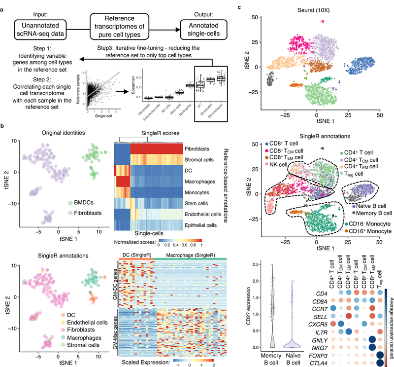
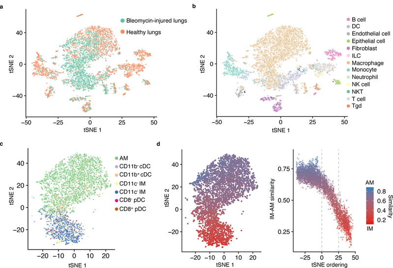
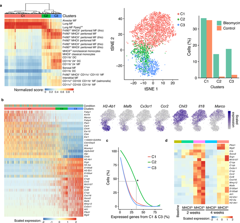
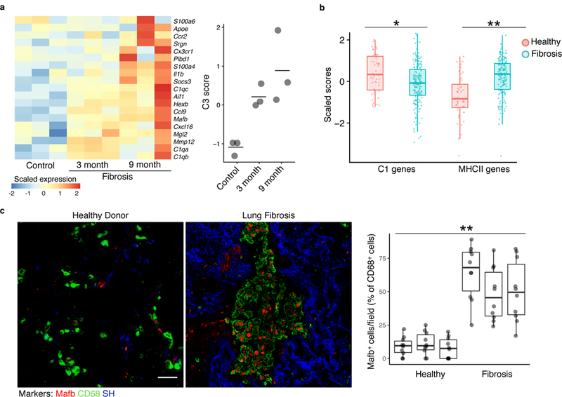
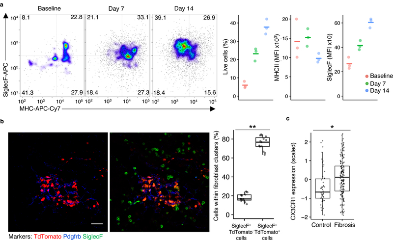
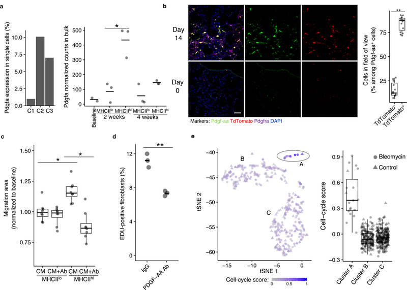
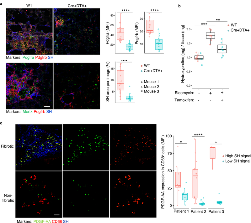
Tissue fibrosis is a major cause of mortality that results from the deposition of matrix proteins by an activated mesenchyme. Macrophages accumulate in fibrosis, but the role of specific subgroups in supporting fibrogenesis has not been investigated in vivo. Here, we used single-cell RNA sequencing (scRNA-seq) to characterize the heterogeneity of macrophages in bleomycin-induced lung fibrosis in mice. A novel computational framework for the annotation of scRNA-seq by reference to bulk transcriptomes (SingleR) enabled the subclustering of macrophages and revealed a disease-associated subgroup with a transitional gene expression profile intermediate between monocyte-derived and alveolar macrophages. These CX3CR1+SiglecF+ transitional macrophages localized to the fibrotic niche and had a profibrotic effect in vivo. Human orthologs of genes expressed by the transitional macrophages were upregulated in samples from patients with idiopathic pulmonary fibrosis. Thus, we have identified a pathological subgroup of transitional macrophages that are required for the fibrotic response to injury.


Figures

Figure 1.
Figure 1.. Reference-based annotation of scRNA-seq.
a, Schematic of SingleR, a protocol for cell-type annotation by reference to transcriptomes of pure cell types. b, tSNE plot of a published scRNA-seq dataset of fibroblast and bone marrow-derived dendritic cells (top left). Heatmap of SingleR scores for top correlated cell types (top right). SingleR annotation of cell identity indicated on t-SNE plot (bottom left). Expression in single cells of genes derived by differential expression analysis of published microarray data from bone marrow-derived, GM-CSF-cultured macrophages (GM-Macs) and DCs (GM-DCs) (bottom right). c, t-SNE plots of a published scRNA-seq dataset of PBMC analyzed by Seurat without (top) and with (middle) SingleR annotations indicated for individual cells. Cell type markers expressed by SingleR annotation (bottom). TCM = central memory T cell; TEM=effector memory T cell.
Figure 2.
Figure 2.. Reference datasets reveal heterogeneity of macrophage subtypes in lung fibrosis.
a-c, t-SNE plots of single-cell suspensions from mouse whole lung sequenced by Drop-seq color-coded by condition (a); annotated by SingleR using the ImmGen database as reference (b); and annotated by SingleR with lung-specific myeloid datasets, (c). Data shown pool replicates (n=3 biologically independent mice for bleomycin-injured and n=6 biologically independent mice for healthy control). d, Quantification of similarity in gene expression of individual cells to bulk RNA-seq profiles of alveolar macrophages (AM) and CD11c+ interstitial macrophages (IM).
Figure 3.
Figure 3.. Hierarchical clustering by reference annotation reveals a transitional macrophage in fibrosis.
a, Heatmap of SingleR annotation scores derived by reference to the ImmGen database with clusters superimposed on the t-SNE plot. MF = macrophages. b, Heatmap of genes differentially expressed between C1 and C3 with examples superimposed on the t-SNE plot. Cells are ordered by the number of non-zero C3 genes. c, Percentage of cells in each cluster that express genes in b (differentially expressed between C1 and C3). Asterisk indicates that 45% of C2 cells express at least 33% of C1 genes and at least 33% of C3 genes. d, Heatmap of the expression of C3 genes in bulk RNA-seq of alveolar macrophages (SiglecF+CD11c+) sorted on MHCIIlo at baseline and both MHCIIlo and MHCIIhi at two time points after bleomycin lung injury (n=2 mice biologically independent mice for baseline, n=3 biologically independent mice at 2 weeks, and n=3 biologically independent mice at 4 weeks).
Figure 4.
Figure 4.. Markers of macrophages identified by scRNA-seq are found in multiple fibrosis models.
a, Heatmap of C3 genes from bleomycin-induced fibrosis measured by microarray in an epithelial telomere dysfunction model of progressive lung fibrosis (n=3 biologically independent mice in each condition, center value is mean). b, Gene set enrichment analysis scores of human orthologues of cluster C1 genes and of MHCII genes in bulk RNA-seq samples from patient lung biopsy specimens. Points indicate individual microarray samples from n=167 and n=50 patients and controls, respectively. c, Immunofluorescence of human fibrotic and healthy control lung (representative images are shown; quantitation is for n=3 independent fibrotic patient lung and n=3 independent healthy lung samples, 10 images per sample). Scale bar, 50 μm. SH=second harmonic imaging of collagen. Box plot center line is median, box limits are upper and lower quartiles, and whiskers denote the largest and smallest values no more than 1.5 times the interquartile range from the limits. Wilcoxon test 2-tailed p-values are presented. * p<0.01, ** p<0.0001.
Figure 5.
Figure 5.. Transitional macrophages expressing CX3CR1, MHCII, and SiglecF localize to sites of fibroblast accumulation after lung injury.
a, Flow cytometry of TdTomato+ lung cells in Cx3cr1-CreERT2 / Rosa26-loxp-STOP-loxp-TdTomato mice with tamoxifen administration before and after injury followed by flow cytometry, with representative pseudocolor plots and values plotted for TdTomato+ cells at three different time points (n=3 biologically independent mice per group, mean is marked). b, Lung immunofluorescence at 14 days after injury in Cx3cr1-CreERT2 / Rosa26-loxp-STOP-loxp-TdTomato mice treated as in a (representative images are shown; quantitation is for n=3 biologically independent mice, 5 images per mouse). Scale bar, 50 μm. c, Expression of CX3CR1 in bulk RNA-seq samples from patient lung biopsy specimens. Box plot center lines are median, box limits are upper and lower quartiles, and whiskers denote the largest and smallest values no more than 1.5 times the interquartile range from the limits. Wilcoxon test 2-sided p-values are presented. * p <0.001; ** p < 0.0001.
Figure 6.
Figure 6.. Pdgf-aa is a macrophage-derived trophic factor for fibroblasts.
a, Pdgfa expression in lung scRNA-seq macrophage clusters and by bulk RNA-seq of SiglecF+CD11c+ lung macrophages after bleomycin injury. Mean is marked. Wald test 2-sided p-value is presented (for the lung scRNA-seq data, n=3 biologically independent mice for bleomycin-injured and n=6 biologically independent mice for healthy control; for the bulk RNA-seq data, n=2 biologically independent mice for baseline, n=3 biologically independent mice for all other conditions). b, Lung immunofluorescence at baseline (bottom) and 14 days after injury (top) in tamoxifen-induced Cx3cr1-CreERT2 / Rosa26loxpSTOPloxp-TdTomato mice (representative images are shown; quantitation is for the 14-day time point in n=3 biologically independent mice, 5 images per mouse). Scale bar, 50 μm. c, 3T3 fibroblast gap closure in response to conditioned media (CM) from lung macrophages sorted by MHCII expression, with and without Pdgf-aa blocking antibody (Ab; n=8 biologically independent mice total, each point a separate assay). d, EDU quantitation in 3T3 fibroblasts co-cultured with TdTomato+ cells sorted from tamoxifen-induced Cx3cr1-CreERT2 / Rosa26-loxp-STOP-loxp-TdTomato mice 14 days after injury. Mean is marked (n=3 biologically independent co-cultures). e, t-SNE of SingleR-annotated fibroblasts from the lung scRNA-seq dataset showing intensity of cell cycle signature (n=3 biologically independent mice for bleomycin, n=6 biologically independent mice for control). Box plot center lines are median, box limits are upper and lower quartiles, and whiskers denote the largest and smallest values no more than 1.5 times the interquartile range from the limits. Wilcoxon test 2-sided p-values are presented. * p < 0.01; ** p < 0.0001.
Figure 7.
Figure 7.. Cx3cr1-derived macrophages drive fibroblast accumulation and fibrosis after lung injury.
a, Lung immunofluorescence of fibroblast and macrophage markers 21 days after bleomycin injury in Cx3cr1-CreERT2 / Rosa26-loxp-STOP-loxp-Diphtheria Toxin A and wild-type mice (quantitation is for n=3 biologically independent mice per group, 5 images per mouse). b, Fibrosis measured by hydroxyproline assay for lung collagen content in mice treated as in a (blue points are Cx3cr1-CreERT2 / Rosa26loxpSTOPloxp-Diptheria Toxin A mice and red points are wild type; n=10, n=7, and n=7 biologically independent mice per group from left to right). Scale bar, 50 μm. c, Immunofluorescence of fibrotic (high SH) and non-fibrotic (low SH) regions from explanted human fibrotic lung, with quantification of individual images from 3 separate patients (8 high and 9 low regions in patient 1, 16 high and 16 low in patient 2, and 3 high and 6 low in patient 3). SH=second harmonic signal for collagen. Scale bar, 50 μm. Box plot center lines are median, box limits are upper and lower quartiles, and whiskers denote the largest and smallest values no more than 1.5 times the interquartile range from the limits. Wilcoxon test 2-sided p-values are presented. * p < 0.05; ** p < 0.01; *** p < 0.001; **** p < 0.0001.

Similar articles

Cited by

References

    1. Rockey DC, Bell PD & Hill JA Fibrosis--A Common Pathway to Organ Injury and Failure. N Engl J Med 373, 96, doi:10.1056/NEJMc1504848.(2015). - DOI - PubMed
    1. Blackwell TS et al. Future directions in idiopathic pulmonary fibrosis research. An NHLBI workshop report. Am J Respir Crit Care Med 189, 214–222, doi:10.1164/rccm.201306–1141WS.(2014). - DOI - PMC - PubMed
    1. Rock JR et al. Multiple stromal populations contribute to pulmonary fibrosis without evidence for epithelial to mesenchymal transition. Proc Natl Acad Sci U S A 108, E1475–1483, doi:10.1073/pnas.1117988108. (2011). - DOI - PMC - PubMed
    1. El Agha E et al. Two-Way Conversion between Lipogenic and Myogenic Fibroblastic Phenotypes Marks the Progression and Resolution of Lung Fibrosis. Cell Stem Cell 20, 261–273 e263, doi:10.1016/j.stem.2016.10.004. (2017). - DOI - PMC - PubMed
    1. Hung C et al. Role of lung pericytes and resident fibroblasts in the pathogenesis of pulmonary fibrosis. Am J Respir Crit Care Med 188, 820–830, doi:10.1164/rccm.201212–2297OC. (2013). - DOI - PMC - PubMed

Publication types

MeSH terms

Substances