. 2019 Feb;25(2):263-269.
doi: 10.1038/s41591-018-0296-z. Epub 2019 Jan 14.

Biomimetic 3D-printed scaffolds for spinal cord injury repair

Affiliations

Biomimetic 3D-printed scaffolds for spinal cord injury repair

Jacob Koffler et al. Nat Med. 2019 Feb.

Abstract

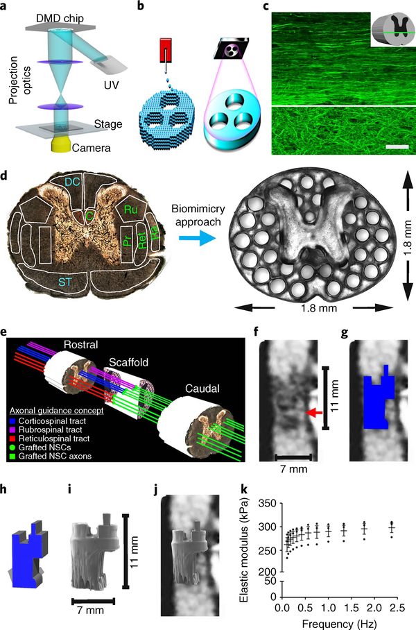
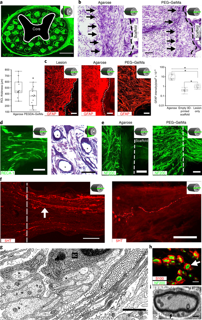
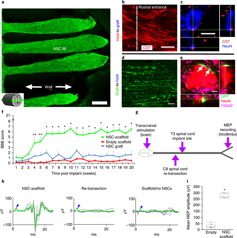
Current methods for bioprinting functional tissue lack appropriate biofabrication techniques to build complex 3D microarchitectures essential for guiding cell growth and promoting tissue maturation1. 3D printing of central nervous system (CNS) structures has not been accomplished, possibly owing to the complexity of CNS architecture. Here, we report the use of a microscale continuous projection printing method (μCPP) to create a complex CNS structure for regenerative medicine applications in the spinal cord. μCPP can print 3D biomimetic hydrogel scaffolds tailored to the dimensions of the rodent spinal cord in 1.6 s and is scalable to human spinal cord sizes and lesion geometries. We tested the ability of µCPP 3D-printed scaffolds loaded with neural progenitor cells (NPCs) to support axon regeneration and form new 'neural relays' across sites of complete spinal cord injury in vivo in rodents1,2. We find that injured host axons regenerate into 3D biomimetic scaffolds and synapse onto NPCs implanted into the device and that implanted NPCs in turn extend axons out of the scaffold and into the host spinal cord below the injury to restore synaptic transmission and significantly improve functional outcomes. Thus, 3D biomimetic scaffolds offer a means of enhancing CNS regeneration through precision medicine.


Conflict of interest statement

Competing interests

The authors declare no competing interests.

Figures

Fig. 1 |
Fig. 1 |. The 3D-printed scaffold mimics the spinal cord architecture.
a, 3D-printer setup: an ultraviolet light source (365nm wavelength); a computer for sliced image-flow generation and system synchronization; a digital micromirror device (DMD) for optical pattern generation; a set of projection optics; a stage for sample position control; and a CCD (charge-coupled device) imaging system for on-line monitoring of the fabrication process. b, μCPP layerless 3D printing creates structures without discrete layers. In extrusion-based 3D-printing approaches (left), as the stage moves in the z axis and material is extruded, layers are created between the drops. μCPP printing (right) creates a structure with one continuous layer. c, Heavy chain neurofilament (NF200) labeling of axons in intact T3 rat spinal cord. Rostral is to the left, and caudal is to the right of the image. The axons in the white matter (top of the panel) are highly organized into parallel arrays traveling from rostral to caudal. The axons in the gray matter (bottom of the panel) are not linear. The white line demarcates the interface between the white and gray matter. The inset schematic diagram indicates the orientation of the horizontal section. d, Axonal projections in the spinal cord are linearly organized into regions (fascicles) containing axons of related function. Motor systems are shown in green and sensory systems are shown in blue. C, corticospinal tract; Ru, rubrospinal tract; Ra, raphespinal tract; Ret, reticulospinal tract; Pr, propriospinal tract; ST, dpinothalamic tract; DC, dorsal column sensory axons. Our scaffold mimic the linear organization of white matter. Channels are precisely printed in 3D space. e, A schematic diagram explaining the axonal alignment and guidance hypothesis. A lesioned host axon (for example, a corticospinal tract (CST), rubrospinal or reticulospinal axon) regenerates into the scaffold and forms a synapse onto a NPC-derived axon inside a channel, and the NPC neuron in turn extends an axon out of the scaffold below the injury site (green lines) into the same white matter fasciculus below the lesion, guided by a linear microchannel. The scaffold maintains its 3D coordinates across the lesion, matching the natural host architecture. f, A sagittal mid-cervical T1-weighted magnetic resonance image of a human clinically complete (ASIA A) SCI. A sliver of spared host white matter is evident on the anterior aspect (right side) of the lesion (arrow). The size of the lesion is noted next to the image. g, The traced outline of the cystic lesion cavity from f. h, A computer-aided design (CAD) 3D model of the scaffold to be 3D-printed, corresponding to the precise lesion shape. i, The printed scaffold matches the size shown in f. j, A hypothetical fit of the printed 3D scaffold in a human contusion cavity. k, Mechanical measurement of the scaffold elastic modulus using dynamic mechanical analysis. n = 3 scaffold samples tested by dynamic mechanical analysis. The data are presented are means± s.e.m. Scale bar, 50μm (c).
Fig. 2 |
Fig. 2 |. Four weeks in vivo performance of empty 3D-printed scaffold implants.
a, A cross-section through an implanted scaffold labeled for axons (NF200). The inset schematic diagrams indicate the section orientation. b, Nissl stain reveals a reactive cell layer (arrows) at the site of implantation of an agarose scaffold (left) or a PEGDA-GelMa scaffold (right). Rostral is to the left and caudal is to the right. The black dashed line demarcates the interface of the host spinal cord with the scaffold. The box and whisker plot shows the quantification of the mean reactive cell layer (RCL) thickness. The boxes show the 25th-75th percentile range, and the center line is the median. Whiskers show 1.5 times the interquartile range (IQR) from the 25th or 75th percentile values. *P < 0.0019 (Student’s t-test), n = 12 animals. c, A host glial ‘scar’ is revealed by GFAP immunoreactivity in animals with lesion only (no scaffold) (left), an agarose scaffold (middle) or a 3D-printed scaffold (right). Rostral is to the left and caudal is to the right. In the 3D-printed scaffold, the glial processes align longitudinally with the channels. The box and whisker plot shows the quantification of the mean GFAP intensity in the host spinal cord surrounding the lesion site. The boxes show the 25th-75th percentile range, and the center mark is the median. Whiskers show 1.5 times IQR from the 25th or 75th percentile values. *P < 0.0001 (one-tailed ANOVA with post hoc Tukey’s), n = 11 animals. d, Scaffold vascularization shown by RECA-1 immunolabeling (left) and toluidine blue stain (asterisks indicate vessels) (right). e, Left, Host axons (labeled for NF200) do not enter agarose scaffolds. Right, Host axons enter 3D-printed scaffolds. The dashed line indicates the host/scaffold interface on the rostral end of the lesion site. f, Host 5HT-labeled serotonergic axons (white arrow) regenerate into an empty scaffold (left) and reach the caudal end of the channel (right). The white dashed line demarcates the rostral entrance to the channel. g, Electron microscopy within a channel demonstrates axons (asterisks) associated with a neighboring ensheathing Schwann cell (SC). h, A magnified view of a channel in a showing S100-labeled Schwann cells ensheathing NF200-labeled axons (arrow). i, Electron microscopy of a channel demonstrates a myelinated axon in the scaffold with a Schwann cell. Scale bars, 500 μm (a), 200 μm (b), 100 μm (c,e), 25 μm (d, left), 20 μm (d, right), 50 μm (f), 1 μm (g), 5 μm (h), 0.5 μm (i).
Fig. 3 |
Fig. 3 |. Four weeks in vivo performance of NPC-loaded 3D-printed scaffold implants.
a, Channels are filled with GFP-expressIng NPCs. The inset schematic diagram indicates the orientation of the horizontal sections in all panels of this figure. b, The rostral entrance to the channel is penetrated by host axons (labeled for NF200 (NF)); the host axons are distinguished from graft-derived axons by the absence of GFP expression. c, Implanted GFP-expressing NSCs extend linear axons within the scaffold. Rostral is to the left and caudal is to the right. d, 5HT-labeled host serotonergic axons enter the NPC-filled channel from the rostral (left) aspect of the lesion and regenerate linearly in the channel (arrow). e, 5HT-labeled host axons exit the caudal aspect of the channel to regenerate into the host spinal cord distal to the lesion (arrow). The white line demarcates the exit from the caudal channel to the caudal spinal cord. f, 5HT host axons regenerating into scaffold channels form appositional contacts (arrows) with dendrites (MAP2) of implanted NPCs (labeled for GFP). g, Quantification of the mean number of 5HT axons reaching the caudal end of the scaffold (one-tail ANOVA P < 0.0322, post hoc Tukey’s), n = 10 animals. The boxes show the 25th-75th percentile range, and the center mark is the median. Whiskers show 1.5 times IQR from the 25th or 75th percentile values. h, At the ultrastructural level, axons of varying diameters (asterisks) are present within channels and many axons are myelinated (M). i, An ultrastructural image showing an oligodendrocyte (green) sending multiple processes to myelinate and ensheath axons (red). j, Synapses (arrows) form between axons within channels and the dendrites of implanted NPCs. The synapses are asymmetric with presynaptic boutons containing rounded vesicles. Scale bars, 200 μm (a), 50 μm (b), 10 μm (c,f), 100 μm (d,e), 500 nm (h), 0.2 μm (i), 200 nm (j).
Fig. 4 |
Fig. 4 |. Long-term in vivo studies of 3D-printed scaffolds loaded with NPCs.
a-e, Anatomy at 6 months post implant. a, The channels are structurally intact and filled with GFP-expressing NPCs. The inset schematic diagram indicates the orientation of the horizontal sections in all panels of this figure, with rostral to the left. b, Corticospinal axons enter the scaffold and extend linearly in a caudal direction. c, CST axons converge on a NeuN-labeled neuron inside the channel, forming bouton-like contacts with the soma. d, GFP axons extend out from the scaffold into the host white and gray matter caudal to the lesion. Ventrolateral white matter, 2 mm caudal to the lesion. e, NPC-derived GFP-labeled axons form excitatory contacts (VGlut2) on gray matter host neurons (labeled for NeuN) located 2 mm caudal to the lesion (white arrows). f-i, Behavioral studies. f, BBB motor scores after complete transection (repeated-measures ANOVA; **P < 0.0232, *P < 0.0008; mean±s.e.m, n = 10 animals). g, Schematic diagram of the electrophysiology study performed at 6 months post implant. Transcranial electrical stimulation is applied to the motor cortex in the brain and MEPs are recorded from the hindlimbs. h, Rats with 3D-printed, NPC-filled scaffolds exhibit MEP responses that are abolished by subsequent re-transection of the spinal cord immediately above the scaffold. Animals with empty scaffolds show no MEPs. The blue arrowheads mark stimulation artifacts. The green lines represent averages of several individual stimulations shown in black. i, The mean MEP amplitude is significantly greater in animals implanted with NPC-containing scaffolds. The boxes show the 25th-75th percentile range, and the center mark is the median. Whiskers show 1.5 times IQR from the 25th or 75th percentile values. (Student’s t-test; P < 0.0001, n = 10 animals). Scale bars, 250μm (a), 50μm (b), 10μm (c), 40μm (d), 5μm (e).

Comment in

Similar articles

Cited by

References

    1. Kadoya K, et al. Spinal cord reconstitution with homologous neural grafts enables robust corticospinal regeneration. Nat. Med 22, 479–487 (2016). - PMC - PubMed
    1. Lu P et al. Long-distance growth and connectivity of neural stem cells after severe spinal cord injury. Cell 150, 1264–1273 (2012). - PMC - PubMed
    1. NSCISC Annual Statistical Report - Model Systems Public Version (National Spinal Cord Injury Statistical Center, University of Alabama at Birmingham, 2014).
    1. Murphy SV & Atala A 3D bioprinting of tissues and organs. Nat. Biotechnol 32, 773–785 (2014). - PubMed
    1. Soman P, Chung PH, Zhang AP & Chen S Digital microfabrication of user-defined 3D microstructures in cell-laden hydrogels. Biotechnol. Bioeng 110, 3038–3047 (2013). - PMC - PubMed

Publication types

Substances