

Primary Cutaneous Angiosarcoma of the Eyelid: A Diagnostic and Therapeutic Challenge
- PMID: 30643767
- PMCID: PMC6322085
- DOI: 10.1159/000485427
Primary Cutaneous Angiosarcoma of the Eyelid: A Diagnostic and Therapeutic Challenge
Abstract
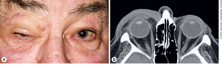
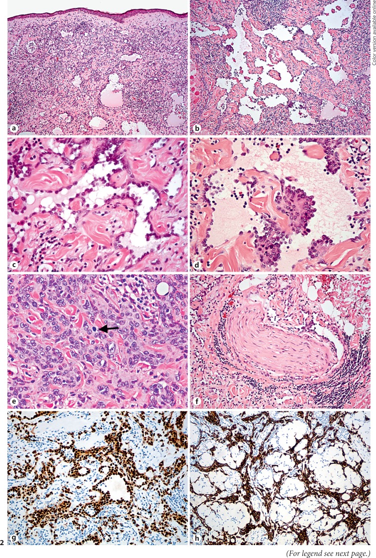
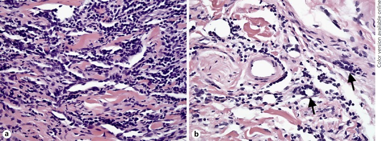
Primary cutaneous angiosarcoma is a rare vasoformative malignant neoplasm that can present a diagnostic and therapeutic challenge. We describe a 76-year-old Caucasian man with right upper eyelid swelling and nodularity, initially suspected clinically to represent either ocular adnexal lymphoma or basal cell carcinoma. Incisional biopsy and wide resection of the mass with frozen section control of margins were interpreted as compatible with hobnail (Dabska-retiform) hemangioendothelioma. Foci of atypia were noted in the tumor, raising speculation of evolution into a more aggressive neoplasm, such as conventional angiosarcoma. The patient subsequently underwent two additional wide resections with frozen section control of margins in an attempt to obtain complete excision of residual tumor, which demonstrated histopathologic features favoring angiosarcoma. The histologic material from the original and subsequent resections was sent in consultation to several soft tissue pathology experts and the final diagnosis of low-grade cutaneous angiosarcoma was established. Despite repeated surgical interventions, there was continued persistence of the tumor in the deep orbital tissues. Various management options, including adjuvant radiotherapy/chemotherapy with and without orbital exenteration, were discussed. The patient decided against further surgical intervention and is currently undergoing adjuvant radiotherapy/chemotherapy. This case illustrates the diagnostic and management difficulties of ocular adnexal angiosarcoma.
Keywords: Angiosarcoma; Eye; Eyelid; Hemangioendothelioma; Hobnail hemangioendothelioma; Retiform hemangioendothelioma; Tumor.
Figures
References
-
- Goldblum JR, Folpe AL, Weiss SW, Malignant vascular tumors . Enzinger and Weiss' Soft Tissue Tumors. In: Goldblum JR, Folpe AL, Weiss SW, editors. 6th ed. Philadelphia: Elsevier Saunders; 2014. pp. pp 703–723.
-
- Hamill EB, Agrawal M, Diwan AH, Winthrop KL, Marx DP. Angiosarcoma of the eyelid with superimposed enterobacter infection. Ophthal Plast Reconstr Surg. 2016;32:e59–e60. - PubMed
-
- Lemanski N, Farber M, Carruth BP, Wladis EJ. Primary adnexal angiosarcoma masquerading as periorbital hematoma. Surv Ophthalmol. 2014;59:655–659. - PubMed
LinkOut - more resources
Full Text Sources
