. 2019 Jan 15;10(1):218.
doi: 10.1038/s41467-018-08211-9.

Structure of tRNA splicing enzyme Tpt1 illuminates the mechanism of RNA 2'-PO4 recognition and ADP-ribosylation

Affiliations

Structure of tRNA splicing enzyme Tpt1 illuminates the mechanism of RNA 2'-PO4 recognition and ADP-ribosylation

Ankan Banerjee et al. Nat Commun. .

Abstract

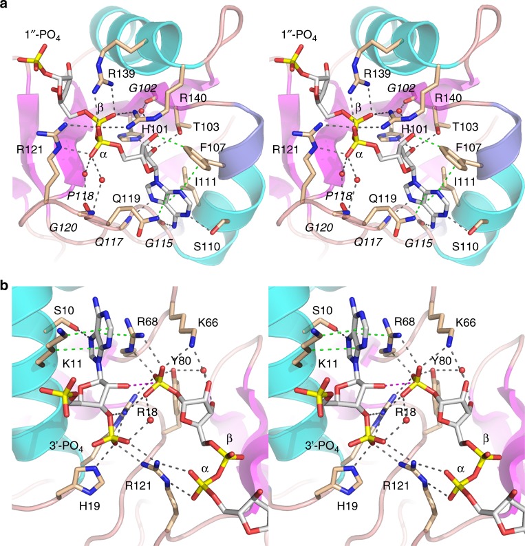
Tpt1 is an essential agent of fungal tRNA splicing that removes the 2'-PO4 at the splice junction generated by fungal tRNA ligase. Tpt1 catalyzes a unique two-step reaction whereby the 2'-PO4 attacks NAD+ to form an RNA-2'-phospho-ADP-ribosyl intermediate that undergoes transesterification to yield 2'-OH RNA and ADP-ribose-1″,2″-cyclic phosphate products. Because Tpt1 is inessential in exemplary bacterial and mammalian taxa, Tpt1 is seen as an attractive antifungal target. Here we report a 1.4 Å crystal structure of Tpt1 in a product-mimetic complex with ADP-ribose-1″-phosphate in the NAD+ site and pAp in the RNA site. The structure reveals how Tpt1 recognizes a 2'-PO4 RNA splice junction and the mechanism of RNA phospho-ADP-ribosylation. This study also provides evidence that a bacterium has an endogenous phosphorylated substrate with which Tpt1 reacts.


Conflict of interest statement

The authors declare no competing interests.

Figures

Fig. 1
Fig. 1
Two-step Tpt1-catalyzed mechanism of 2′-PO4 removal from a 2′-PO4, 3′-5′ phosphodiester RNA junction. The Tpt1 reaction pathway comprises the two chemical steps shown in which: (1) the RNA 2′-PO4 reacts with NAD+ to expel nicotinamide and form a 2′-phospho-ADP-ribosylated RNA intermediate; and (2) transesterification of the ADP-ribose 2′′-OH to the RNA 2′-PO4 displaces the RNA 2′-OH and generates ADP-ribose-1″,2″-cyclic phosphate
Fig. 2
Fig. 2
Overview of the Tpt1 structure and active site ligands. a The primary structure of CthTpt1 is aligned to those of Tpt1 enzymes from Escherichia coli (Eco), Runella slithyformis (Rsl), and Saccharomyces cerevisiae (Sce). Positions of amino acid side chain identity or similarity in all four proteins are indicated by black dots above the CthTpt1 sequence. Gaps in the alignment are indicated by dashes. The constituents of a conserved Arg-His-Arg-Arg tetrad essential for Tpt1 activity are highlighted in gold shading. The secondary structure elements of CthTpt1—magenta β strands, cyan α helices, and blue 310 helices—are displayed above the CthTpt1 sequence. The boundary between the N-terminal RNA lobe (aa 1–85) and the C-terminal NAD+ lobe (aa 86–182) is demarcated by a two-headed arrow below the alignment. Amino acids that contact the ADP-ribose moiety in the NAD+ site are denoted by red dots below the alignment. Amino acids that contact the RNA splice junction (the pAp and the 1″-PO4) are denoted by blue dots. b Stereo view of the CthTpt1 tertiary structure, depicted as a cartoon model with magenta β strands, cyan α helices, and blue 310 helices. The N and C termini are indicated and the α helices are numbered. The ADP-ribose-1″-PO4 and coenzyme A ligands are rendered as a stick models with gray carbons. c A stereo view of the simulated annealing omit 2Fo-Fc map of the ADP-ribose-1″-PO4 and pAp ligands in the active site, contoured at 1σ, is shown in gray mesh. Anomalous difference density for the phosphorus atoms of ADP-ribose-1″-PO4 and pAp, contoured at 3σ, is shown in blue mesh. The simulated annealing omit 2Fo-Fc and anomalous difference density maps were calculated with both ligands omitted from the model. Anomalous peaks for the sulfur atoms of Met60 and Cys94 were apparent at 4σ but are not shown because they are remote from the two active site ligands
Fig. 3
Fig. 3
Tpt1 active site. Stereo views of the atomic interactions of Tpt1 with the ADP-ribose moiety of the ADP-ribose-1″-PO4 product a and the pAp and 1″-PO4 moieties b. Selected Tpt1 amino acids are depicted as stick models with beige carbons. The ligands are depicted as stick models with gray carbons. Waters are depicted as red spheres. Hydrogen bonds are denoted by black dashed lines and van der Waals contacts by green dashed lines. The distance (3.5 Å) from the ribose 2′-O to the 1″-P in b is indicated by a magenta dashed line. Amino acids that make side-chain contacts to the ligand are labeled in plain font; those that make main-chain contacts to the ligands are labeled in italics
Fig. 4
Fig. 4
Surface electrostatics and comparison to ApeTpt1 apoenzyme. a Surface electrostatic model of the CthTpt1 protein generated in Pymol. ADP-ribose-1″-PO4 and CoA ligands are depicted as stick models. The top view highlights positive potential (blue) across the RNA and NAD+ lobes surrounding the ligands, with ADP-ribose-1″-PO4 being encased below an “arch″ formed by two arginines. The bottom view shows the adenine of pAp wedged into a groove on the positive surface. b Surface electrostatic model is shown of the Aeropyrum pernix (Ape) Tpt1 apoenzyme (pdb 1WFX) with ADP-ribose-1″-PO4 imported from the CthTpt1 structure. c Superposition of the CthTpt1 (beige) and ApeTpt1 (cyan) structures. The arginines that form the arch over ADP-ribose-1″-PO4 in CthTpt1 are shown as stick models. Side chain movement of ApeTpt1 Arg 134 versus CthTpt1 Arg139 gives access to the NAD+ binding pocket
Fig. 5
Fig. 5
Homology of the Tpt1 NAD+ lobe to ADP-ribosylating toxins. A side-by-side alignment of the homologous segments of the Tpt1•ADPR-1″-PO4 (beige), Pierisin1•NAD+ (pdb 5H6J, cyan), and diphtheria toxin•NAD+ (pdb 1TOX, green) structures is shown. The ligands are depicted as stick models with gray carbons. Amino acid contacts to the ADP-ribose moieties are indicated by dashed lines
Fig. 6
Fig. 6
Structure-guided mutagenesis. a Reaction mixtures containing 100 mM Tris-HCl (pH 7.5), either 1 mM or 0.1 mM NAD+ as specified, 0.2 µM (2 pmol) 5′ 32P-labeled 6-mer 2′-PO4 RNA (shown at bottom), and 0.01 µM (0.1 pmol) wild-type or mutant RslTpt1 as specified were incubated for 30 min at 37 °C. Tpt1 was omitted from the control reaction mixture in lane –. The reaction products were analyzed urea-PAGE and visualized by autoradiography. The positions of the 6-mer 2′-PO4 RNA substrate (2′-P), 2′-P-ADPR RNA intermediate, and 2′-OH RNA product (2′-OH) are indicated on the right. The extents of 2′-OH RNA product formation (expressed as percent of total labeled RNA) are indicated below the lanes. b tpt1∆ complementation was performed by plasmid shuffle as described under Methods. Serial dilutions of the indicated S. cerevisiae tpt1RslTPT1 strains were spot-tested for growth on YPD agar at the temperatures specified

References

    1. Culver GM, et al. An NAD derivative produced during transfer RNA splicing: ADP-ribose 1″-2″ cyclic phosphate. Science. 1993;261:206–208. doi: 10.1126/science.8392224. - DOI - PubMed
    1. Spinelli SL, Kierzek R, Turner DH, Phizicky EM. Transient ADP-ribosylation of a 2′-phosphate implicated in its removal from ligated tRNA during splicing in yeast. J. Biol. Chem. 1999;274:2637–2644. doi: 10.1074/jbc.274.5.2637. - DOI - PubMed
    1. Steiger MA, Kierzek R, Turner DH, Phizicky EM. Substrate recognition by a yeast 2′-phosphotransferase involved in tRNA splicing and its Escherichia coli homolog. Biochemistry. 2001;40:14098–14105. doi: 10.1021/bi011388t. - DOI - PubMed
    1. Steiger MA, Jackman JE, Phizicky EM. Analysis of 2′-phosphotransferase (Tpt1p) from Saccharomyces cerevisiae: evidence for a conserved two-step reaction mechanism. RNA. 2005;11:99–106. doi: 10.1261/rna.7194605. - DOI - PMC - PubMed
    1. Munir A, Abdullahu L, Damha MJ, Shuman S. Two-step mechanism and step-arrest mutants of Runella slithyformis NAD+-dependent tRNA 2′-phosphotransferase Tpt1. RNA. 2018;24:1144–1157. doi: 10.1261/rna.067165.118. - DOI - PMC - PubMed

Publication types

MeSH terms