. 2019 Jan 15;9(1):138.
doi: 10.1038/s41598-018-36932-w.

Metabolome and transcriptome-wide effects of the carbon storage regulator A in enteropathogenic Escherichia coli

Affiliations

Metabolome and transcriptome-wide effects of the carbon storage regulator A in enteropathogenic Escherichia coli

Volker Berndt et al. Sci Rep. .

Abstract

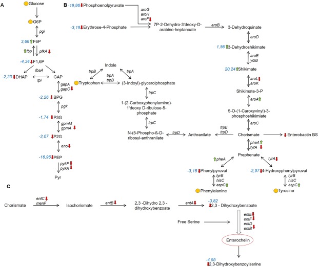
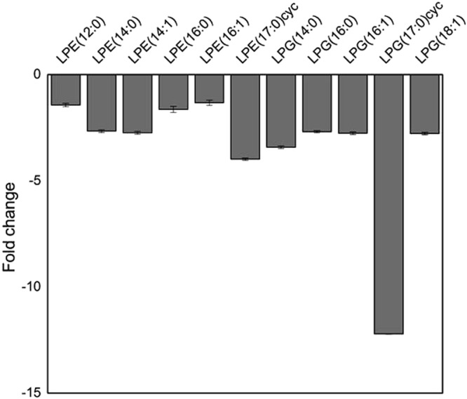
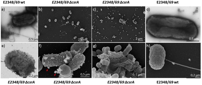
The carbon storage regulator A (CsrA) is a conserved global regulatory system known to control central carbon pathways, biofilm formation, motility, and pathogenicity. The aim of this study was to characterize changes in major metabolic pathways induced by CsrA in human enteropathogenic Escherichia coli (EPEC) grown under virulence factor-inducing conditions. For this purpose, the metabolomes and transcriptomes of EPEC and an isogenic ∆csrA mutant derivative were analyzed by untargeted mass spectrometry and RNA sequencing, respectively. Of the 159 metabolites identified from untargeted GC/MS and LC/MS data, 97 were significantly (fold change ≥ 1.5; corrected p-value ≤ 0.05) regulated between the knockout and the wildtype strain. A lack of csrA led to an accumulation of fructose-6-phosphate (F6P) and glycogen synthesis pathway products, whereas metabolites in lower glycolysis and the citric acid cycle were downregulated. Associated pathways from the citric acid cycle like aromatic amino acid and siderophore biosynthesis were also negatively influenced. The nucleoside salvage pathways were featured by an accumulation of nucleosides and nucleobases, and a downregulation of nucleotides. In addition, a pronounced downregulation of lyso-lipid metabolites was observed. A drastic change in the morphology in the form of vesicle-like structures of the ∆csrA knockout strain was visible by electron microscopy. Colanic acid synthesis genes were strongly (up to 50 fold) upregulated, and the abundance of colanic acid was 3 fold increased according to a colorimetric assay. The findings expand the scope of pathways affected by the csrA regulon and emphasize its importance as a global regulator.


Conflict of interest statement

The authors declare no competing interests.

Figures

Figure 1
Figure 1
(a) CsrA and Tir-Bla expression in different growth media: Western blot analysis of the Tir-ß-lactamase (Tir-Bla) fusion protein from whole cell lysates under different growth conditions at an OD600 of ~1. The growth of EPEC in DMEM under microaerobic conditions was used as a positive control for Tir-Bla synthesis (right). (b) Levels of CsrA in different strains and growth media at an OD600 of ~1 determined by Western blots. Blots were cropped. Full length blots are shown in Fig. S4.
Figure 2
Figure 2
Influence of CsrA on metabolic pathways of EPEC. Regulation of glycolysis (A), aromatic amino acid synthesis (B) and enterochelin synthesis (C) in the EPEC ΔcsrA mutant. Red arrows indicate downregulation, green arrows indicate upregulation, and yellow circles indicate unchanged levels in the ΔcsrA knockout strain compared to the wildtype. Quantitative fold changes and corrected p-values are listed in the Supplementary Dataset S1. Metabolites: glucose-6-phosphate G6P, fructose-6-phosphate F6P, fructose 1,6-bisphosphate F1,6P, dihydroxyacetonphosphate DHAP, glyceraldehyde-3-phosphate GAP, bisphosphoglycerate BGP, 3-phosphoglycerate 3PGA, 2-phosphglycerate 2PGA, phosphoenolpyruvate PEP, pyruvate pyr. Fold changes of significant regulated metabolites are shown in blue. Genes for enzymes: Glucose-6-phosphate isomerase pgi, fructose-1,6-bisphosphatase fbp, Phosphofructokinase pfkA, fructose bisphosphate aldolase fbaA, triose phosphate isomerase tpi, glyceraldehyde 3-phosphate dehydrogenase-A gapA, glyceraldehyde 3-phosphate dehydrogenase C gapC, Phosphoglycerate kinase pgk, Phosphoglycerate mutase M gpmM, Phosphoglycerate mutase A gpmA, enolase eno, pyruvate kinase I pykF, pyruvate kinase II pykA, 2,3-Dihydro-2,3-dihydroxybenzoate dehydrogenase entA, Asochorismatase entB, Isochorismate synthase entC, Enterochelin synthase component D entD, Component of 2,3-dihydroxybenzoate-AMP ligase entE, Enterochelin synthase component F entF, Component of isochorismate synthase menF, 3-deoxy-D-arabino-heptulosonate-7-phosphate synthase aroG, Component of 2-dehydro-3-deoxyphosphoheptonate aldolase aroH/aroF, 3-Dehydroquinate synthase aroB, 3-Dehydroquinate dehydratase aroD, Shikimate 5-dehydrogenase aroE, shikimate dehydrogenase ydiB, shikimate kinase II aroL, shikimate kinase I aroK, 5-enolpyruvyl shikimate-3-phosphate synthase aroA, chorismate synthase aroC, bifunctional chorismate/prephenate dehydratase pheA/tyrA, anthranilate synthase component trpE/D, bifunctional: N-(5-phosphoribosyl)anthranilate isomerase and indole-3-glycerolphosphate synthase trpC, Component of histidinol-phosphate aminotransferase hisC, Aspartate aminotransferase aspC.
Figure 3
Figure 3
Bar diagram of lysolipid levels of EPEC. Fold changes in abundance for lysoPE and lysoPG species in the ΔcsrA strain compared to the EPEC wild type. Data represent averages from positive and negative measurements. Only significant values were used to create this figure (corrected p-value ≤ 0.05). Error bars represent the average relative error of the fold change. All log2fold changes and corrected p-values are listed in Table S3.
Figure 4
Figure 4
Phenotypic changes of the ΔcsrA mutant. Transmission electron microscopy (TEM) pictures of negative stained EPEC (E2348/69) wildtype (a,d) and the isogenic E2348/69 ΔcsrA mutant (e). Field electron microscopy (FEM) pictures of EPEC (E2348/69)(b,h) and the E2348/69 ΔcsrA mutant (c,f,g).

References

    1. Rothbaum R, McAdams AJ, Giannella R, Partin JC. A clinicopathologic study of enterocyte-adherent Escherichia coli: a cause of protracted diarrhea in infants. Gastroenterology. 1982;83:441–454. - PubMed
    1. Rothbaum RJ, Partin JC, Saalfield K, McAdams AJ. An ultrastructural study of enteropathogenic Escherichia coli infection in human infants. Ultrastruct. Pathol. 1983;4:291–304. doi: 10.3109/01913128309140582. - DOI - PubMed
    1. Knutton S, Baldini MM, Kaper JB, McNeish AS. Role of plasmid-encoded adherence factors in adhesion of enteropathogenic Escherichia coli to HEp-2 cells. Infect. Immun. 1987;55:78–85. - PMC - PubMed
    1. Finlay BB, Rosenshine I, Donnenberg MS, Kaper JB. Cytoskeletal composition of attaching and effacing lesions associated with enteropathogenic Escherichia coli adherence to HeLa cells. Infect. Immun. 1992;60:2541–2543. - PMC - PubMed
    1. Alikhani MY, Mirsalehian A, Fatollahzadeh B, Pourshafie MR, Aslani MM. Prevalence of enteropathogenic and shiga toxin-producing Escherichia coli among children with and without diarrhoea in Iran. J. Health Popul. Nutr. 2007;25:88–93. - PMC - PubMed

MeSH terms