. 2019 Sep;235(3):543-554.
doi: 10.1111/joa.12931. Epub 2019 Jan 15.

Neuroserpin expression during human brain development and in adult brain revealed by immunohistochemistry and single cell RNA sequencing

Affiliations

Neuroserpin expression during human brain development and in adult brain revealed by immunohistochemistry and single cell RNA sequencing

Istvan Adorjan et al. J Anat. 2019 Sep.

Abstract

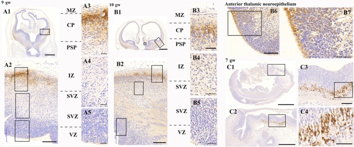
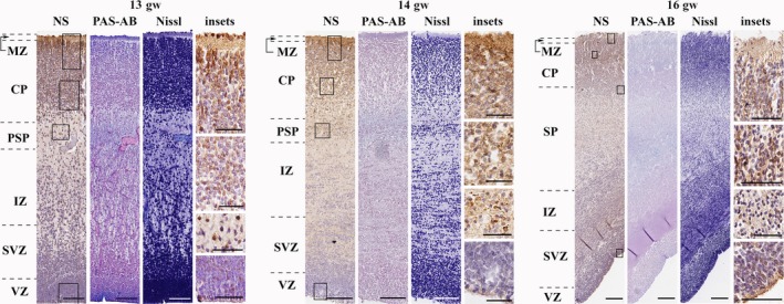
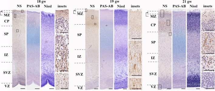
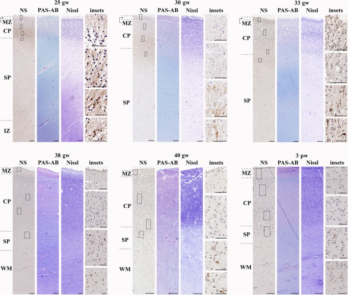
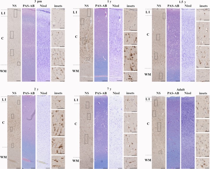
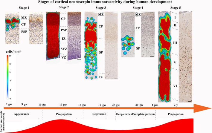
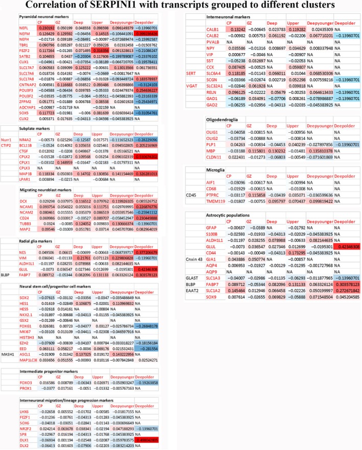
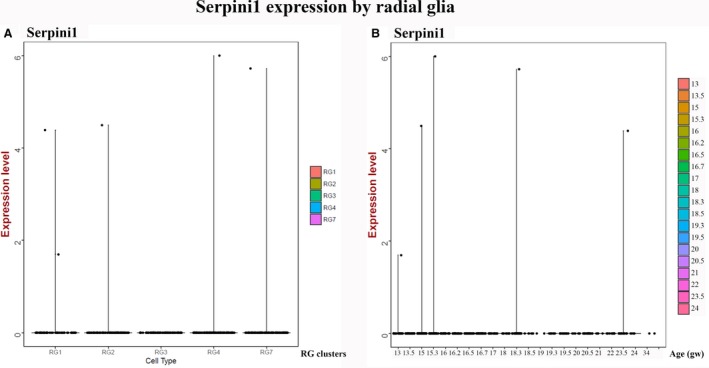
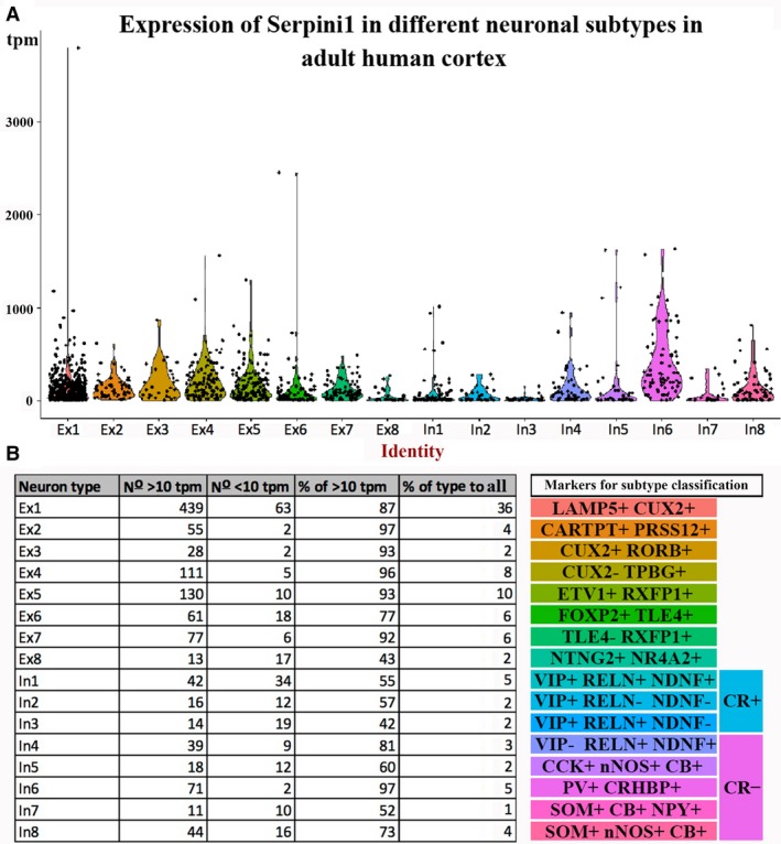
Neuroserpin is a serine-protease inhibitor mainly expressed in the CNS and involved in the inhibition of the proteolytic cascade. Animal models confirmed its neuroprotective role in perinatal hypoxia-ischaemia and adult stroke. Although neuroserpin may be a potential therapeutic target in the treatment of the aforementioned conditions, there is still no information in the literature on its distribution during human brain development. The present study provides a detailed description of the changing spatiotemporal patterns of neuroserpin focusing on physiological human brain development. Five stages were distinguished within our examined age range which spanned from the 7th gestational week until adulthood. In particular, subplate and deep cortical plate neurons were identified as the main sources of neuroserpin production between the 25th gestational week and the first postnatal month. Our immunohistochemical findings were substantiated by single cell RNA sequencing data showing specific neuronal and glial cell types expressing neuroserpin. The characterization of neuroserpin expression during physiological human brain development is essential for forthcoming studies which will explore its involvement in pathological conditions, such as perinatal hypoxia-ischaemia and adult stroke in human.

Keywords: human brain; neurodevelopment; neuroserpin; subplate.


Conflict of interest statement

The authors have no conflicts of interest to declare.

Figures

Figure 1
Figure 1
Neuroserpin expression in the first trimester of human brain development. A1–A5: neuroserpin is expressed in the upper cortical plate of the ventrolateral pallium in the 9th gw. B1–B5: neuroserpin is expressed in the upper cortical plate of the ventrolateral pallium in the 10th gw. B6–B7: the anterior thalamic neuroepithelium also contained neuroserpin‐ip cells with long processes. C1–C4: neuroserpin is expressed by a distinct neuronal population in the rhombencephalon in the 7th gw. No telencephalic expression was observed at this stage of development. MZ, marginal zone; CP, cortical plate; PSP, pre‐subplate; IZ, intermediate zone; SVZ, subventricular zone; VZ, ventricular zone. Scale bars: (A1) 1000 μm, (A2, B2) 200 μm, (A3–A5) B3–B6, (C3) 100 μm, (B1, C1) 2000 μm, (B7, C4) 50 μm, (C2) 500 μm.
Figure 2
Figure 2
Neuroserpin expression in the early second trimester (frontal lobe). Neuroserpin‐ip cells were present through the cortical plate and subplate. They had long processes and elongated perikarya showing migratory morphology. Minimal expression was seen in the marginal zone. Neuroserpin‐ip cells were observed in the ventricular zone of the lateral and caudal ganglionic eminences. MZ, marginal zone; CP, cortical plate; PSP, pre‐subplate; SP, subplate; IZ, intermediate zone; SVZ, subventricular zone; VZ, ventricular zone. Scale bars: (13th gw) NS, PASAB, Nissl: 100 μm, insets: 50 μm; (14th–16th gw) NS, PASAB, Nissl: 200 μm, insets: 50 μm.
Figure 3
Figure 3
Neuroserpin expression in the middle second trimester in the human telencephalon (frontal lobe). Neuroserpin‐ip cells with elongated perikarya and long processes directed to the pial surface were mainly seen in the middle and deep cortical plate and subplate. No neuroserpin‐ip cells were observed in the upper cortical plate. MZ, marginal zone; CP, cortical plate; SP, subplate; IZ, intermediate zone; SVZ, subventricular zone; VZ, ventricular zone. Scale bars: (18th gw) NS, PASAB, Nissl: 100 μm; (19th–21st gw) NS, PASAB, Nissl: 200 μm, insets: 50 μm.
Figure 4
Figure 4
Neuroserpin expression from the late second trimester until the first postnatal month (frontal lobe). Neuroserpin‐ip cells were situated to the deep cortical plate and subplate with a pyramidal and elongated shape. The upper and middle regions of the cortical plate were devoid of neuroserpin immunoreactivity. MZ, marginal zone; CP, cortical plate; SP, subplate; IZ, intermediate zone; WM, white matter. Scale bars: (25th gw–3rd pw) NS, PASAB, Nissl: 200 μm, insets: 50 μm.
Figure 5
Figure 5
Neuroserpin expression from the third postnatal month until adulthood in the cortex (frontal lobe). Neuroserpin‐ip neurons were observed throughout layers of the cortex with the exception of layer 1, where minimal immunoreactivity was present. The majority of neuroserpin‐ip neurons had a pyramidal morphology. LI, layer 1; C, cortical layers 2–6; WM, white matter. Scale bars: (3 pm–adult) NS, PASAB, Nissl: 200 μm, insets: 50 μm.
Figure 6
Figure 6
Heatmaps showing the characteristic stages of neuroserpin immunoreactivity in the developing human cortex based on the density of neuroserpin‐ip cells. MZ, marginal zone; CP, cortical plate; PSP, pre‐subplate; IZ, intermediate zone; SVZ, subventricular zone; VZ, ventricular zone. Scale bars: (stage 1) 50 μm, (stage 2) 80 μm, (stage 3) 130 μm, (stage 4) 140 μm, (stage 5) 150 μm.
Figure 7
Figure 7
Correlation levels of Serpini1 to different transcripts during development visualised by heatmap (based on Nowakowski et al. 2017). Transcripts are grouped in specific cell types. CP, cortical plate; GZ, granular zone; Deep, deep cortical; Upper, upper cortical; Deep younger, deep cortical <15 gestational week; Deep older, deep cortical > 15th gw.
Figure 8
Figure 8
Expression of neuroserpin in developing human cortex described by single cell transcriptomics analysis. (A) Violin plot for neuroserpin expression in different radial glial subtypes (counts per million). (B) Violin plot for neuroserpin expression by radial glial clusters plotted along the age (gestational week) (Nowakowski et al. 2017).
Figure 9
Figure 9
Expression of neuroserpin in adult human temporal cortex as identified by single cell transcriptomics analysis. (A) Violin plot for neuroserpin expression in each subtype of excitatory and inhibitory neurons from dataset of single cell RNA sequencing data in Lake et al. (2016). (B) Table summarizing the number and percentage of neurons for each subtype that expressed neuroserpin above the threshold (tpm > 10). Subtype‐markers are shown on the right hand side.

References

    1. Adorjan I, Ahmed B, Feher V, et al. (2017) Calretinin interneuron density in the caudate is lower in autism spectrum disorder. Brain 140, 2028–2040. - PMC - PubMed
    1. Bannister LH, Berry MM, Collins P, et al. (eds) (1995) Gray's Anatomy, 38th edn, p. 344. New York: Churchill Livingstone.
    1. Chéret J, Lebonvallet N, Misery L, et al. (2012) Expression of neuroserpin, a selective inhibitor of tissue‐type plasminogen activator in the human skin. Exp Dermatol 21, 710–720. - PubMed
    1. Dillman AA, Majounie E, Ding J, et al. (2017) Transcriptomic profiling of the human brain reveals that altered synaptic gene expression is associated with chronological aging. Sci Rep 7, 1–12. - PMC - PubMed
    1. Eyre JA, Miller S, Clowry GJ, et al. (2000). Functional corticospinal projections are established prenatally in the human foetus permitting involvement in the development of spinal motor centres. Brain 123(Pt 1), 51–64. - PubMed

Publication types