. 2019 Jan 30;141(4):1555-1567.
doi: 10.1021/jacs.8b10992. Epub 2019 Jan 15.

Biophysical Characterization of a Disabled Double Mutant of Soybean Lipoxygenase: The "Undoing" of Precise Substrate Positioning Relative to Metal Cofactor and an Identified Dynamical Network

Affiliations

Biophysical Characterization of a Disabled Double Mutant of Soybean Lipoxygenase: The "Undoing" of Precise Substrate Positioning Relative to Metal Cofactor and an Identified Dynamical Network

Shenshen Hu et al. J Am Chem Soc. .

Abstract

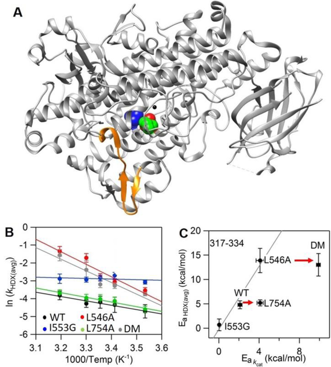
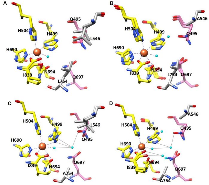
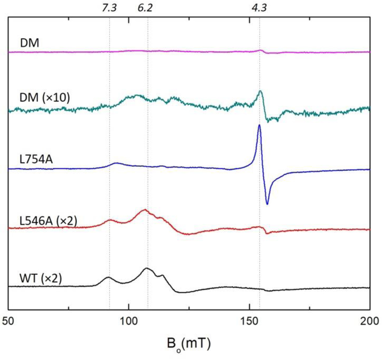
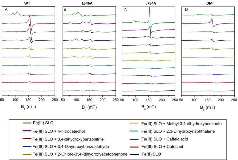
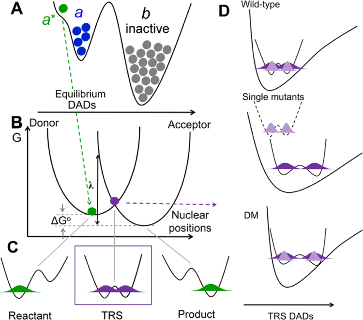
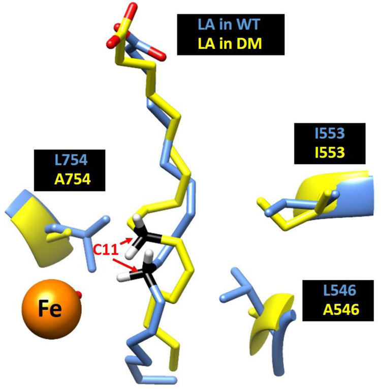
Soybean lipoxygenase (SLO) has served as a prototype for understanding the molecular origin of enzymatic rate accelerations. The double mutant (DM) L546A/L754A is considered a dramatic outlier, due to the unprecedented size and near temperature-independence of its primary kinetic isotope effect, low catalytic efficiency, and elevated enthalpy of activation. To uncover the physical basis of these features, we herein apply three structural probes: hydrogen-deuterium exchange mass spectrometry, room-temperature X-ray crystallography and EPR spectroscopy on four SLO variants (wild-type (WT) enzyme, DM, and the two parental single mutants, L546A and L754A). DM is found to incorporate features of each parent, with the perturbation at position 546 predominantly influencing thermally activated motions that connect the active site to a protein-solvent interface, while mutation at position 754 disrupts the ligand field and solvation near the cofactor iron. However, the expanded active site in DM leads to more active site water molecules and their associated hydrogen bond network, and the individual features from L546A and L754A alone cannot explain the aggregate kinetic properties for DM. Using recently published QM/MM-derived ground-state SLO-substrate complexes for WT and DM, together with the thorough structural analyses presented herein, we propose that the impairment of DM is the combined result of a repositioning of the reactive carbon of linoleic acid substrate with regard to both the iron cofactor and a catalytically linked dynamic region of protein.


Figures

Figure 1:
Figure 1:
Peptide 317–334 (in orange) remote from the active site exhibits mutation-induced changes in the enthapic barrier for HDX rates (EaHDX(avg)) in L546A, I553G and DM but not in L754A. (A) The SLO structure with highlighted active site residues L546 (in green), L754 (in blue) and I553 (in red). In panel B, Arrhenius-like plots of the weighted average exchange rate, (ln(kHDX(avg)) for WT, I553G, L546A, L754A and DM are compared. In panel C, the relationship between EaHDX(avg) and the enthalpic barrier for hydrogen tunneling (Ea(kcat)) is represented. The solid line is the trend among I553G, WT and L546A previously reported. The bottom arrow indicates that L754A shows no difference in EaHDX(avg) relative to WT. The top arrow shows that there is no further difference in EaHDX(avg) when L754A is further introduced into L546A.
Figure 2:
Figure 2:
Coordination geometry of the iron center of (A) WT SLO (PDB: 5T5V); (B) L546A (PDB:5TQN); (C) L754A (PDB:5TR0); and (D) DM (PDB: 5TQO). Oxygen atoms are colored in red, nitrogens in blue, iron center in orange and water in cyan. Carbons in first sphere ligands are colored in yellow, second-sphere ligands in pink, side chains on position 546 and 754 in grey. Bonds between iron and first-sphere ligands (yellow) are drawn as dashed grey lines, and hydrogen-bonding interactions between newly added water in the active site with the first sphere and second sphere ligands are shown as solid grey lines. There are no significant differences in side chain conformers for SLO variants based on the electron densities analysis.
Figure 3:
Figure 3:
EPR spectra (X-band, 6.5K) of oxidized WT, L546A, L754A and DM SLO (140 μM) in 0.1 M potassium phosphate pH = 7.0 buffer. The peak intensities of L546A, L754A and DM have been corrected for the iron content of each mutant: WT (0.9), L546A (0.50), L754A (0.70), DM (0.70). A 10-x amplified EPR spectrum of Fe(III) DM SLO is presented for comparison. Expanded EPR spectra are shown in Figure S5.
Figure 4:
Figure 4:
EPR spectra after Fe(III)-SLOs are anaerobically incubated with catechol ligands in 0.1 M potassium phosphate pH = 7.0 buffer at 4°C for 12 hrs: (A) WT; (B) L546A; (C) L754A; (D) DM. The EPR spectra of oxidized Fe(III) SLO vs Fe(II) SLO are shown at the top and bottom of each panel for comparison.
Figure 5.
Figure 5.
Rate constants for enzyme catalyzed nonadiabatic hydrogen transfer can be formalized as kobs = KeqkPCET., , (A) Keq (<< 1) represents a stochastic ground state search through inactive conformations (b) to reach catalytically active E-S complexes (a conformer). Subsequent thermal sampling further reduces the distance between the H-donor and acceptor as seen in the pre-tunneling a* conformer. (B-D) illustrate contributions from kPCET: (B) Heavy atom protein motions produce transiently degenerate energies for the reactant and product wells, a prerequisite for wave function overlap at the tunneling ready state (TRS) (C). (D) The effective potential along the DAD sampling coordinate varies, starting with ΔEa ≅ 0 for native enzyme (top), becoming ΔEa >>0 when the DAD is elongated following single mutations (middle) and finally arriving at the catastrophic DM scenario in which the DAD is elongated, but coupled with a rigid DAD sampling potential (bottom). In Frame D, light purple refers to the shorter wave function distribution for deuterium, and dark purple represents the more distributive protium wave function.
Figure 6:
Figure 6:
A comparison between the ground state substrate binding mode in WT (Blue) and DM (yellow). The reactive carbon C11 is colored as black, iron colored as orange.
Scheme 1.
Scheme 1.
Reaction mechanism for the first and rate limiting step of the stereospecific oxidation of linoleic acid catalyzed by SLO, where the hydrogen atom transfer from substrate LA to ferric hydroxide cofactor occurs by a PCET process.
Scheme 2.
Scheme 2.
The free energy profile for WT (red) is represented by a two state thermodynamic model involving E-S and the enzyme-bound pentadienyl intermediate, E-I. In the case of DM, the most stable E-S complex (Figure 6) must first isomerize to a pre-tunneling configuration (E-S’) that converts via PCET to the mispositioned pentadienyl intermediate (E-I’) with a driving force similar to WT. The return of the bound intermediate E-I’ to a more stable binding position completes the reaction. The dashed blue line has been added as a possible model for the QM/MM computation.

References

    1. Radzicka A; Wolfenden R, A Proficient Enzyme. Science 1995, 267, 90–93. - PubMed
    1. Edwards DR; Lohman DC; Wolfenden R, Catalytic Proficiency: The Extreme Case of S-O Cleaving Sulfatases. J. Am. Chem. Soc 2012, 134, 525–531. - PubMed
    1. Bruice TC, A View at the Millennium: The Efficiency of Enzymatic Catalysis. Acc. Chem. Res 2002, 35, 139–148. - PubMed
    1. Benkovic SJ; Hammes-Schiffer S, A Perspective on Enzyme Catalysis. Science 2003, 301, 1196–1202. - PubMed
    1. Nagel ZD; Klinman JP, A 21st Century Revisionist’s View at a Turning Point in Enzymology. Nat. Chem. Biol 2009, 5, 543–550. - PubMed

Publication types