

Biophysical stimulation of bone and cartilage: state of the art and future perspectives
- PMID: 30645684
- PMCID: PMC6399199
- DOI: 10.1007/s00264-018-4274-3
Biophysical stimulation of bone and cartilage: state of the art and future perspectives
Abstract
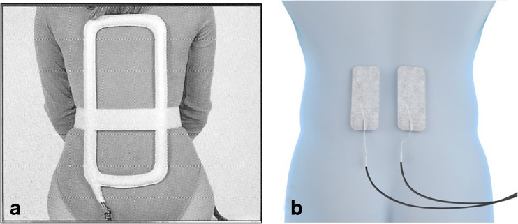
Introduction: Biophysical stimulation is a non-invasive therapy used in orthopaedic practice to increase and enhance reparative and anabolic activities of tissue.
Methods: A sistematic web-based search for papers was conducted using the following titles: (1) pulsed electromagnetic field (PEMF), capacitively coupled electrical field (CCEF), low intensity pulsed ultrasound system (LIPUS) and biophysical stimulation; (2) bone cells, bone tissue, fracture, non-union, prosthesis and vertebral fracture; and (3) chondrocyte, synoviocytes, joint chondroprotection, arthroscopy and knee arthroplasty.
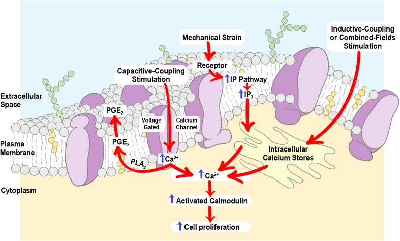
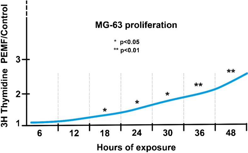
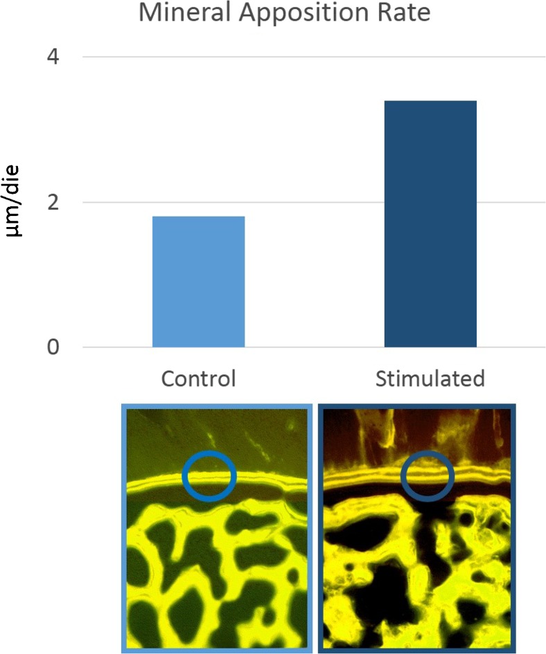
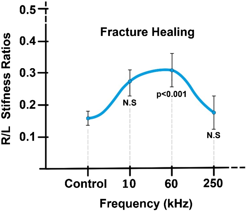
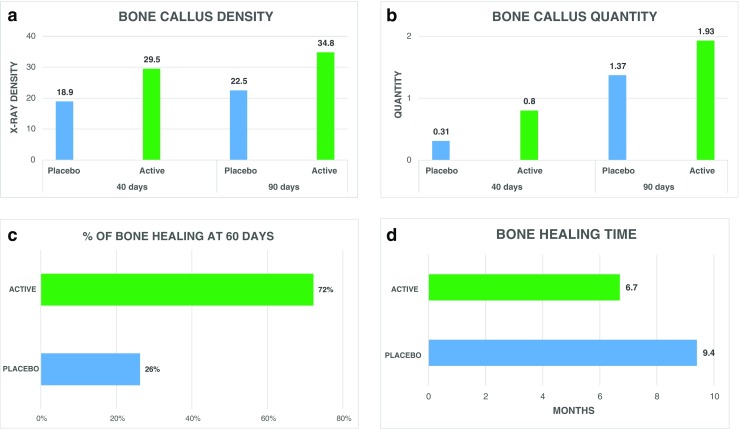
Results: Pre-clinical studies have shown that the site of interaction of biophysical stimuli is the cell membrane. Its effect on bone tissue is to increase proliferation, synthesis and release of growth factors. On articular cells, it creates a strong A2A and A3 adenosine-agonist effect inducing an anti-inflammatory and chondroprotective result. In treated animals, it has been shown that the mineralisation rate of newly formed bone is almost doubled, the progression of the osteoarthritic cartilage degeneration is inhibited and quality of cartilage is preserved. Biophysical stimulation has been used in the clinical setting to promote the healing of fractures and non-unions. It has been successfully used on joint pathologies for its beneficial effect on improving function in early OA and after knee surgery to limit the inflammation of periarticular tissues.
Discussion: The pooled result of the studies in this review revealed the efficacy of biophysical stimulation for bone healing and joint chondroprotection based on proven methodological quality.
Conclusion: The orthopaedic community has played a central role in the development and understanding of the importance of the physical stimuli. Biophysical stimulation requires care and precision in use if it is to ensure the success expected of it by physicians and patients.
Keywords: Biophysical stimulation; Bone tissue; CCEF; Cartilage; LIPUS; PEMF.
Conflict of interest statement
The authors declare that they have no conflict of interest.
Figures
References
-
- Zhou J, Wang JQ, Ge BF, et al. Different electromagnetic field waveforms have different effects on proliferation, differentiation and mineralization of osteoblasts in vitro. Bioelectromagnetics. 2014;35(1):30–38. - PubMed
-
- Clark CC, Wang W, Brighton CT. Up-regulation of expression of selected genes in human bone cells with specific capacitively coupled electric fields. J Orthop Res. 2014;32(7):894–903. - PubMed
-
- Brighton CT, Okereke E, Pollack SR, Clark CC (1992) In vitro bone-cell response to a capacitively coupled electrical field. The role of field strength, pulse pattern, and duty cycle. Clin Orthop Relat Res (285):255–62 - PubMed
-
- De Mattei M, Caruso A, Traina GC, et al. Correlation between pulsed electromagnetic fields exposure time and cell proliferation increase in human osteosarcoma cell lines and human normal osteoblast cells in vitro. Bioelectromagnetics. 1999;20(3):177–182. - PubMed
-
- Leung KS, Cheung WH, Zhang C, Lee KM, Lo HK (2004) Low intensity pulsed ultrasound stimulates osteogenic activity of human periosteal cells. Clin Orthop Relat Res (418):253–9 - PubMed
