

Inadvertent Migration of Umbilical Venous Catheters Often Leads to Malposition
- PMID: 30645997
- PMCID: PMC6518856
- DOI: 10.1159/000494369
Inadvertent Migration of Umbilical Venous Catheters Often Leads to Malposition
Abstract
Background: Migration of umbilical venous catheters (UVCs) has been described anecdotally.
Objectives: The aim of this paper was to investigate migration of UVCs using ultrasonography (US).
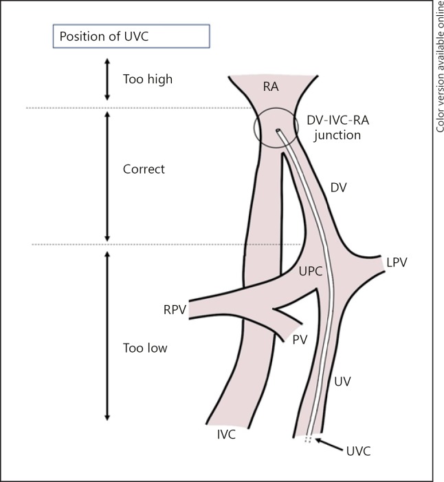
Methods: In a prospective observational study, the position of UVCs was determined using serial US within 24 h, at midweek, and at the end of the week after umbilical catheterization. Migration was recorded in distance and direction. Malposition was defined as a position of the UVC in the heart (right atrium or more distal along the UVC-route), umbilicoportal confluence, or in the umbilical vein. UVC position determined by US was compared with chest X-rays (CXRs) when these were performed for standard care within the same period of 1 h.
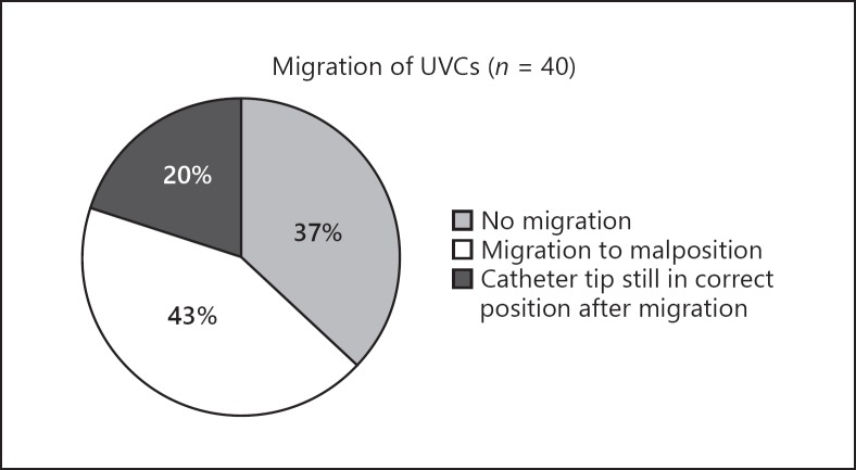
Results: Migration of UVCs was detected with US in 25/40 infants (63%) in 32 occasions, leading to malposition in 17/25 (68%) infants. UVCs migrated inwards in 18/32 (56%), leading to a position within the heart in 17/18 occasions. Most migrations occurred before Day 3 (21/32 [66%]). When a CXR was taken at the same time as US was performed (30 occasions), the assessment of the catheter-tip position differed in 23% of the occasions. When malposition was detected by US, this was detected on routinely performed CXRs in 11% of the occasions.
Conclusions: UVCs often migrate following insertion, often leading to malposition. Awareness for this is needed, and US is a feasible alternative for detecting malposition compared to CXRs and avoids additional radiation. Re-evaluation of the position of UVCs at least once, but within 24 h after placement, is recommended.
Keywords: Malposition; Migration; Umbilical venous catheters.
© 2019 The Author(s) Published by S. Karger AG, Basel.
Figures
References
-
- Hogan MJ. Neonatal vascular catheters and their complications. Radiol Clin North Am. 1999 Nov;37((6)):1109–25. - PubMed
-
- Rand T, Kirchner L, Puig S, Ponhold W, Vergesslich K, Imhof H. [“Lines and tubes” in neonatal intensive care patients] Radiologe. 2000 Jan;40((1)):52–7. - PubMed
-
- Simanovsky N, Ofek-Shlomai N, Rozovsky K, Ergaz-Shaltiel Z, Hiller N, Bar-Oz B. Umbilical venous catheter position: evaluation by ultrasound. Eur Radiol. 2011 Sep;21((9)):1882–6. - PubMed
-
- Hermansen MC, Hermansen MG. Intravascular catheter complications in the neonatal intensive care unit. Clin Perinatol. 2005 Mar;32((1)):141–56. - PubMed
-
- Anderson J, Leonard D, Braner DA, Lai S, Tegtmeyer K. Videos in clinical medicine. Umbilical vascular catheterization. N Engl J Med. 2008 Oct;359((15)):e18. - PubMed
Publication types
MeSH terms
LinkOut - more resources
Full Text Sources
