Review
. 2019 Jan 14;20(2):303.
doi: 10.3390/ijms20020303.

The Interaction between Viral and Environmental Risk Factors in the Pathogenesis of Multiple Sclerosis

Affiliations
Review

The Interaction between Viral and Environmental Risk Factors in the Pathogenesis of Multiple Sclerosis

Rachael Eugenie Tarlinton et al. Int J Mol Sci. .

Abstract

Multiple sclerosis (MS) is a chronic debilitating inflammatory disease of unknown ethology targeting the central nervous system (CNS). MS has a polysymptomatic onset and is usually first diagnosed between the ages of 20⁻40 years. The pathology of the disease is characterized by immune mediated demyelination in the CNS. Although there is no clinical finding unique to MS, characteristic symptoms include sensory symptoms visual and motor impairment. No definitive trigger for the development of MS has been identified but large-scale population studies have described several epidemiological risk factors for the disease. This list is a confusing one including latitude, vitamin D (vitD) levels, genetics, infection with Epstein Barr Virus (EBV) and endogenous retrovirus (ERV) reactivation. This review will look at the evidence for each of these and the potential links between these disparate risk factors and the known molecular disease pathogenesis to describe potential hypotheses for the triggering of MS pathology.

Keywords: Epstein Barr Virus (EBV); HERV; central nervous system (CNS); cytotoxic T lymphocytes (CTL); multiple sclerosis.


Conflict of interest statement

The authors declare no conflict of interest

Figures

Figure 1
Figure 1
Magnetic resonance imaging (MRI) of MS and health brain. (A) Typical MS lesions in the right optical nerve on coronal T2 image; (B,C) Typical MS lesions in periventricular white matter on axials T2 and T1 post contrast images, respectively; (DF) Typical MS lesions in the white matter of sub-tentorial structures (pons, right middle cerebellar peduncle): on axials T2 and T1-post-contrast, and coronal T2 images, respectively; (G,H) MRI of the healthy adult brain: normal sub-tentorial structures on axial T2 images, normal periventricular white matter on axial FLAIR images, respectively.
Figure 2
Figure 2
Clinical course of MS. The course of MS can be divided into clinically isolated syndrome (CIS) and clinically defined RPMS. Radiologically isolated syndrome (RIS) is defined by incidental imaging findings suggesting inflammatory demyelination in the absence of clinical signs or symptoms, while CIS is identified as the first clinical presentation of the disease. SPMS is diagnosed retrospectively by a history of gradual worsening after an initial relapsing disease course [53].
Figure 3
Figure 3
Global risk of development of MS. The prevalence of MS increases further from the equator in either hemisphere. Prevalence is higher in North America and Europe (291 and 232 per 100,000 respectively) and lowest in South America, Sub-Saharan Africa and East Asia, at 58 per 100,000 respectively [88]. It appears that highest incidence rate is registered among patients residing above the 42° latitude.
Figure 4
Figure 4
Pathogenesis of MS.

References

    1. Popescu B.F., Pirko I., Lucchinetti C.F. Pathology of multiple sclerosis: Where do we stand? Continuum (Minneap Minn) 2013;19:901–921. doi: 10.1212/01.CON.0000433291.23091.65. - DOI - PMC - PubMed
    1. Horowitz A.L., Kaplan R.D., Grewe G., White R.T., Salberg L.M. The ovoid lesion: A new MR observation in patients with multiple sclerosis. AJNR Am. J. Neuroradiol. 1989;10:303–305. - PMC - PubMed
    1. Lucchinetti C., Bruck W., Parisi J., Scheithauer B., Rodriguez M., Lassmann H. Heterogeneity of multiple sclerosis lesions: Implications for the pathogenesis of demyelination. Ann. Neurol. 2000;47:707–717. doi: 10.1002/1531-8249(200006)47:6<707::AID-ANA3>3.0.CO;2-Q. - DOI - PubMed
    1. Calabrese M., Favaretto A., Martini V., Gallo P. Grey matter lesions in MS: From histology to clinical implications. Prion. 2013;7:20–27. doi: 10.4161/pri.22580. - DOI - PMC - PubMed
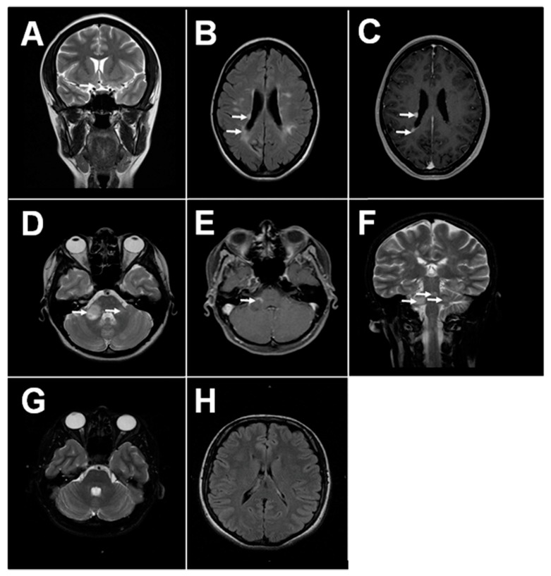
    1. Calabrese M., De Stefano N., Atzori M., Bernardi V., Mattisi I., Barachino L., Morra A., Rinaldi L., Romualdi C., Perini P., et al. Detection of cortical inflammatory lesions by double inversion recovery magnetic resonance imaging in patients with multiple sclerosis. Arch. Neurol. 2007;64:1416–1422. doi: 10.1001/archneur.64.10.1416. - DOI - PubMed

MeSH terms