Review
. 2019 Jan 14;12(2):253.
doi: 10.3390/ma12020253.

Materials for the Spine: Anatomy, Problems, and Solutions

Affiliations
Review

Materials for the Spine: Anatomy, Problems, and Solutions

Brody A Frost et al. Materials (Basel). .

Abstract

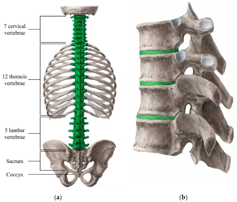
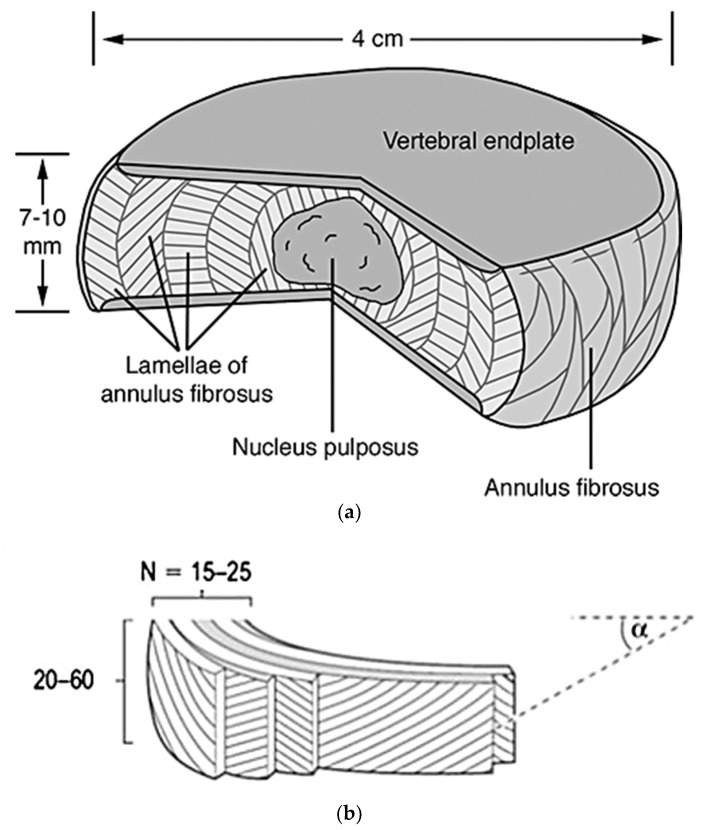
Disc degeneration affects 12% to 35% of a given population, based on genetics, age, gender, and other environmental factors, and usually occurs in the lumbar spine due to heavier loads and more strenuous motions. Degeneration of the extracellular matrix (ECM) within reduces mechanical integrity, shock absorption, and swelling capabilities of the intervertebral disc. When severe enough, the disc can bulge and eventually herniate, leading to pressure build up on the spinal cord. This can cause immense lower back pain in individuals, leading to total medical costs exceeding $100 billion. Current treatment options include both invasive and noninvasive methods, with spinal fusion surgery and total disc replacement (TDR) being the most common invasive procedures. Although these treatments cause pain relief for the majority of patients, multiple challenges arise for each. Therefore, newer tissue engineering methods are being researched to solve the ever-growing problem. This review spans the anatomy of the spine, with an emphasis on the functions and biological aspects of the intervertebral discs, as well as the problems, associated solutions, and future research in the field.

Keywords: degenerative disc disease; herniated disc; intervertebral disc; spinal anatomy; spinal fusion; tissue engineering; total disc replacement.


Conflict of interest statement

The authors declare no conflict of interest.

Figures

Figure 1
Figure 1
Overview of the vertebral column with each specific section labeled for clarification (a). The green highlighted section refers to the part of the spine that contain individual vertebrae, as well as intervertebral discs (IVD). The structure of the vertebrae and IVD (green highlighted) have been added for better visualization (b) [4].
Figure 2
Figure 2
Pictured (a) is a cut out portion of a normal disc depicting the nucleus pulposus, vertebral endplates, and annulus fibrosus. The chosen intervertebral disc is 4 cm wide and 7–10 mm thick [37]. Depicted in the lower image (b) is a diagram showing the detailed structure of the annulus fibrosus, with its 15–25 lamellae comprised of 20–60 collagen fiber bundles. Also shown, is the angle α, which correlates to the directionality of the fibers’ bundles in relation to the vertebrae [38].
Figure 3
Figure 3
Construction of fibrillary collagen as described above [45].
Figure 4
Figure 4
Fluorescence microscopic images of stained components in the outer annulus fibrosus (a), inner annulus fibrosus (b), and nucleus pulposus (c). The microfibrils in relation to cell distribution (blue) and collagen fiber organization (red) indicates the organization of the microfibrils within the ECM of the outer annulus fibrosus. Opposite however, the microfibrils (red) and elastin fibers (green) in the inner annulus fibrosus do not demonstrate any organization or co-localization to any great degree within the ECM. These two distinct characteristics of organization give rise to the varying mechanical properties of each, Section 2.2.1, (3). The microfibrils (red) show a tendency to hover/organize around the nucleus pulposus cells (blue), while the elastin fibers (green) have a tendency to stay dispersed through the entire ECM [51].
Figure 5
Figure 5
The connection of the hyaline cartilage vertebral endplate to the perforated cortical bone of the vertebral body and collagen fibers of the annulus and nucleus. The arrows in the figure refer to the direction of nutrients and blood flow through the different components of the disc, mainly coming from the bone through the vertebral endplates [37].
Figure 6
Figure 6
(a) Schematic representation of the multiple longer and thicker vascular channels throughout the intervertebral disc on a 10-month old female; while (b) represents the vascular channels throughout the disc of a 50-year old adult, showing the retraction and thinning of the channels [37].
Figure 7
Figure 7
The innervation of a healthy intervertebral disc, showing the sinuvertebral nerves and rami communicantes extending into the vertebral foramen and the outer annulus of the disc [37].
Figure 8
Figure 8
A healthy, normal intervertebral disc on the left, shows a distinct difference between the swollen, softer looking nucleus and the ringed annulus. However, during growth and skeletal maturation, the boundary between these components becomes less obvious, and with the nucleus generally becoming more fibrotic and less gel-like, like the highly degenerate disc on the right [79].
Figure 9
Figure 9
MRI scans showing the different grades of disc degeneration based on the Pfirrmann grading system, (IV) referring to Grades (IV): I is representative of Grade (I) degeneration, (II) is representative of Grade (II) degeneration, (III) is representative of Grade (III) degeneration, (IV) is representative of Grade (IV) degeneration, and (V) is representative of Grade (V) degeneration [88].
Figure 10
Figure 10
Sagittal computerized axial tomography (CT scan) image of the cervical spine showing large anterior osteophytes (indicted by the arrows) extending from C5 to C7, which affect the intervertebral disc space [95].
Figure 11
Figure 11
MRI image showing a slight bulge of the annulus into the spinal canal without severe impingement (a). MRI image showing a full lumbar disc herniation with substantial spinal stenosis and nerve-root compression (b) [97].
Figure 12
Figure 12
Example image of spinal fusion surgery using titanium cages loaded with hydroxyapatites and pedicle screws and rods to keep stability and anatomic alignment in spinal segment [114].

References

    1. Britannica T.E.o.E., editor. Encyclopaedia Britannica. Encyclopaedia Britannica, Inc.; Chicago, IL, USA: 2014. Vertebral Column.
    1. Kibler W.B., Press J., Sciascia A. The role of core stability in athletic function. Sports Med. 2006;36:189–198. doi: 10.2165/00007256-200636030-00001. - DOI - PubMed
    1. Agur A.M.R., Dalley A.F. Grant’s Atlas of Anatomy. 12th ed. Lipincott Williams & Wilkins; Pennsylvania, PA, USA: 2009. p. 841.
    1. Vertebral Column. [(accessed on 14 April 2016)]; Available online: https://www.kenhub.com/en/start/c/vertebral-column.
    1. Bogduk N., Mercer S. Biomechanics of the cervical spine. I: Normal kinematics. Clin. Biomech. 2000;15:633–648. doi: 10.1016/S0268-0033(00)00034-6. - DOI - PubMed

LinkOut - more resources