Case Reports
. 2019 Jan 15;19(1):18.
doi: 10.1186/s12886-019-1035-z.

Intravitreal ranibizumab or conbercept for retinal arterial macroaneurysm: a case series

Affiliations
Case Reports

Intravitreal ranibizumab or conbercept for retinal arterial macroaneurysm: a case series

Zhongjing Lin et al. BMC Ophthalmol. .

Abstract

Background: There is no consensus for the standard treatment of retinal arterial macroaneurysm (RAM). Intravitreal anti-vascular endothelium growth factor (anti-VEGF) is an alternative treatment option for RAM. The purpose of this study is to describe the clinical efficacy of intravitreal ranibizumab or intravitreal conbercept for retinal arterial macroaneurysm.

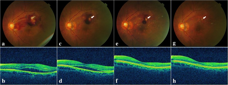
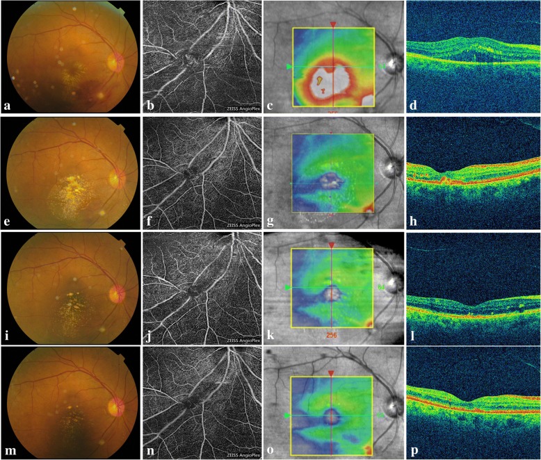
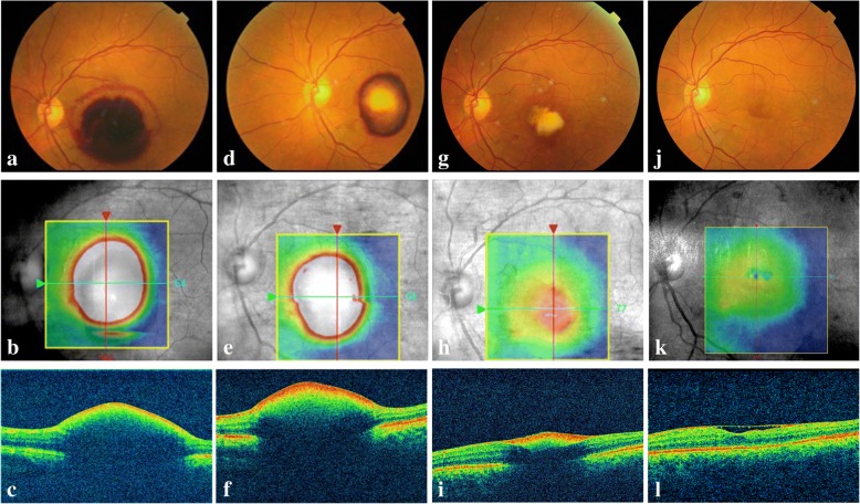
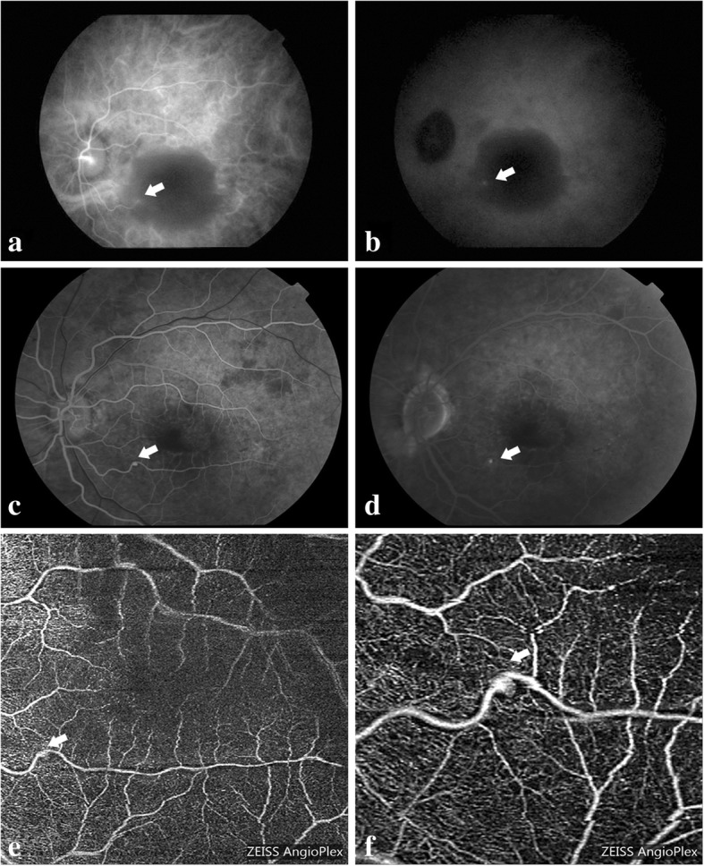
Case presentation: Three cases that presented with symptomatic RAM were treated with intravitreal anti-VEGF agents. Two eyes received two intravitreal ranibizumab injections with a time interval of one month and completed a one-year follow-up, while one eye only received one intravitreal conbercept injection and was followed up for six months. Both the retinal thickness and the visual acuity were significantly improved at the final clinic visit. The macular hemorrhage and edema were resolved. There were no ocular or systemic side effects.

Conclusions: Intravitreal ranibizumab or conbercept might be used as a therapeutic option for symptomatic retinal arterial macroaneurysm patients. Anti-VEGF therapy should be further investigated in a larger series with longer follow-up for this disease profile.

Keywords: Conbercept; Ranibizumab; Retinal arterial macroaneurysm.


Conflict of interest statement

Ethics approval and consent to participate

The study was adhered to the tenets of the Declaration of Helsinki. Written informed consents were obtained from the patients before the surgery. The study conforms to the ethical guidelines of the Ethics Committee of Ruijin Hospital, Shanghai Jiaotong University (Shanghai, China).

Consent for publication

Written informed consents for publication of the clinical details were obtained from the patients.

Competing interests

The authors declare that they have no competing interests.

Publisher’s Note

Springer Nature remains neutral with regard to jurisdictional claims in published maps and institutional affiliations.

Figures

Fig. 1
Fig. 1
Multimodal imaging of the affected eye in case 1. a. Fundus photography showing the supra-temporal hemorrhage and white lesion above the macula at the baseline visit. b. Corresponding OCT scan through the fovea showing SRD at the baseline visit. c. One month after the first injection, fundus photography showing the diminished hemorrhage site and dominant white fibrosis (arrow). d. Corresponding OCT scan through the fovea showing SRD. e. One month after the second injection, fundus photography showing the further diminished hemorrhage site and dominant white fibrosis (arrow). f. Corresponding OCT scan through the fovea showing total resolution of SRD. g. Fundus photography showing no hemorrhage and only white fibrosis left (arrow) at the one-year follow up. h. Corresponding OCT scan through the fovea showing normal appearance of the macular
Fig. 2
Fig. 2
Multimodal imaging of the affected eye in case 2. a. Fundus photography showing infra-temporal RAM with surrounding stellate-shaped exudates involving the fovea at the baseline visit. b. OCT angiography showing the heliciform capillary mass in the RAM in the superficial layer at the baseline visit. c. Corresponding image showing the OCT B-scan lines. d. OCT scan showing SRD in the macular area at the baseline visit. e. Fundus photography showing the absorption of the hemorrhage and the remaining white fibrosis and hard exudates involving the fovea one month after the first injection. f. OCT angiography showing the regression of the heliciform capillary mass in the RAM in the superficial layer one month after the first injection. g. Corresponding image showing the OCT B-scan lines. h. OCT scan showing disappeared SRD in the macular area one month after the first injection. i. Fundus photography showing resolved hard exudates one month after the second injection. j. OCT angiography showing the further regression of the heliciform capillary mass in the RAM in the superficial layer one month after the second injection. k. Corresponding image showing the OCT B-scan lines. l. CT scan through the fovea showing total resolution of SRD one month after the second injection. m. Fundus photography showing no hemorrhage and only white fibrosis (arrow) in the inferior-temporal artery at the one-year follow up. n. OCT angiography showing the absence of the capillary mass at the one-year follow up. o. Corresponding image showing the OCT B-scan lines. p. OCT scan showing normal macular appearance without SRD at the one-year follow up
Fig. 3
Fig. 3
Multimodal imaging of the affected eye in case 2. a. FA image at the early phase showing fluorescein filling of the macroaneurysm (arrow) at the baseline visit. b. FA image at the late phase showing mild leakage (arrow) at the baseline visit. c. FA image at the early phase showing resolution of the macroaneurysm leaving focal perivascular fibrosis (arrow) at the one-year follow up. d. FA image at the late phase showing resolution of the macroaneurysm leaving focal perivascular fibrosis (arrow) at the one-year follow up
Fig. 4
Fig. 4
Multimodal imaging of the affected eye in case 3. a. Color photography at baseline showing the macular hemorrhage involving the posterior pole. b. Corresponding image showing the OCT B-scan lines. c. OCT through the fovea showing the preretinal hemorrhage and increased CMT at baseline visit. d. Color photography showing the partial absorption of the hemorrhage one month after the first injection. e. Corresponding image showing the OCT B-scan lines. f. OCT scan showing the decreasing CMT one month after the first injection. g. Color photography showing the further absorption of the hemorrhage at the 3-month follow-up. h. Corresponding image showing the OCT B-scan lines. i. OCT scan showing further decreased CMT at the 3-month follow-up. j. Color photography at the 6-month follow-up showing almost no hemorrhage and a red lesion at the inferior temporal artery. k. Corresponding image showing the OCT B-scan lines. l. OCT scan at 6-month follow-up showing almost no SRD
Fig. 5
Fig. 5
Multimodal imaging of the affected eye in case 3. a. ICGA at the early phase showing hyperfluoresence at the infratemperal area of the edge of the dark area at the baseline visit. b. ICGA at the late phase showing the continuing lighting in the same area at the baseline visit. c. FA at the early phase showing fluorescein filling of the macroaneurysm at the 6-month follow-up. d. FA at the late phase showing not faded fluorescein filling of the macroaneurysm at the 6-month follow-up. e. OCT-A 6 mm × 6 mm superficial slab revealed the RAM at the 6-month follow-up. f. OCT-A 3 mm × 3 mm superficial slab clearly delineated the RAM at the 6-month follow-up. White arrow indicated the macroaneurysm

References

    1. Robertson DM. Macroaneurysms of the retinal arteries. Trans Am Acad Ophthalmol Otolaryngol. 1973;77(1):55–67. - PubMed
    1. Addel-Khalek MN, Richardson J. Retinal macroaneurysm: natural history and guidelines for treatment. Br J Ophthalmol. 1986;70(1):2–11. doi: 10.1136/bjo.70.1.2. - DOI - PMC - PubMed
    1. Rabb MF, Gagliano DA, Teske MP. Retina arterial macroaneurysms. Surv Ophthalmol. 1988;33(2):73–96. doi: 10.1016/0039-6257(88)90160-9. - DOI - PubMed
    1. McCabe CM, Flynn HW, Jr, Mclean WC, et al. Nonsurgical management of macular hemorrhage secondary to retinal artery macroaneurysms. Arch Ophthaolmol. 2000;118(6):780–785. doi: 10.1001/archopht.118.6.780. - DOI - PubMed
    1. Contreras JE, Mittra RB, Mieler WF, Pollack JS. Retina arterial macroaneurysms. In: Yanoff M, Duker JS, editors. Ophthalmology. 2. Philadelphia: Mosby Elsevier; 2004. pp. 912–917.

Publication types

MeSH terms