

Cervical spine alignment following surgery for adolescent idiopathic scoliosis (AIS): a pre-to-post analysis of 81 patients
- PMID: 30646880
- PMCID: PMC6334400
- DOI: 10.1186/s12893-019-0471-2
Cervical spine alignment following surgery for adolescent idiopathic scoliosis (AIS): a pre-to-post analysis of 81 patients
Abstract
Background: Several studies have emphasized the importance of restoring thoracic kyphosis (TK) in the setting of AIS, but very few have discussed changes in cervical spine alignment following surgery. Aim of this study was to evaluate reciprocal cervical alignment change after modification of global and regional thoracolumbar alignment with surgery in the setting of adolescent idiopathic scoliosis (AIS).
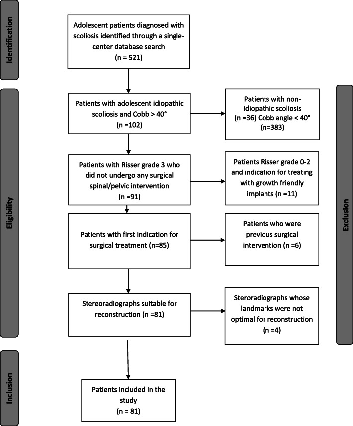
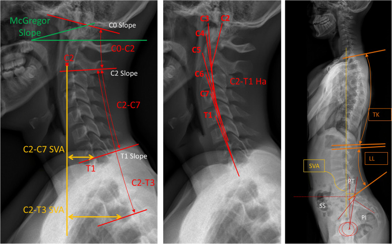
Methods: Baseline and 2-yrs follow-up radiographs of AIS patients (n = 81) were analysed measuring cervical parameters (upper cervical: C2-C0, McGregor Slope; lower cervical: C2-C7, C2-C7 sagittal vertical axis (SVA), C2-T3, C2-T3SVA, C2-T1Harrison (C2-T1Ha), T1 Slope (T1S)), thoracic, lumbar, pelvic and global alignment parameters. Post-operatively, patients were grouped twice; based on changes in TK and SVA. Cervical alignment was compared between groups. Pearson correlation was conducted to examine the relationship between changes in TK, SVA, and cervical alignment.
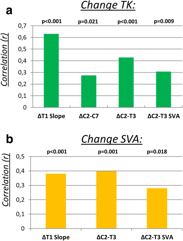
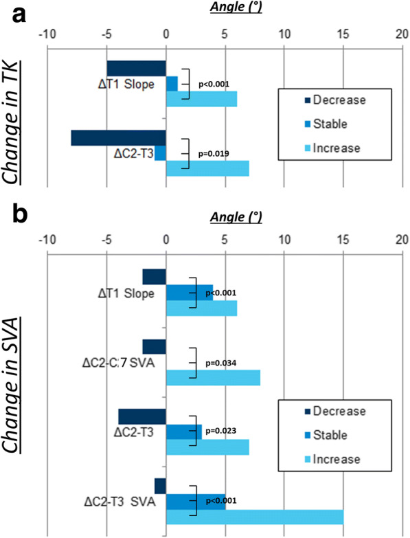
Results: Stratification by change in TK, revealed significant alteration of lower cervical alignment T1S [p < 0.001]), C2-T3 [p = 0.019], C2-T1Ha [p = 0.043]), but there was no reciprocal change in the upper cervical spine. Stratification by SVA revealed a significant coexisting change in the lower cervical spine (T1S [p < 0.001], C2-C7SVA [p = 0.034], C2-T3 [p = 0.023], C2-T3SVA [p = 0.001]). SVA change was not associated to a change in the upper cervical spine. The correlation analysis showed that with a post-operative increase in TK, the cervical spine became more lordotic. Changes in TK were significantly correlated with: ΔT1S, ΔC2-C7, ΔC2-T3, and ΔC2-T3SVA. Similarly, increased cervical kyphosis was found when SVA was decreased post-operatively. Furthermore, there was a significant correlation between change of SVA and both ΔC2-T3 and ΔC2-T3SVA.
Conclusions: In surgically treated AIS patients, changes in global and regional alignment of the thoracolumbar and cervical spinal segments exhibit interdependence. Thus, surgical planning with regard to sagittal deformity in AIS patients should account for the post-operative impact on cervical alignment.
Keywords: AIS; Adolescent scoliosis; Cervical alignment; Cervical spine; Deformity.
Conflict of interest statement
The authors declare that they have no competing interests.
Figures
References
-
- Hey HW, Lau ET, Wong CG, Tan KA, Liu GK, Wong HK. Cervical Alignment Variations in Different Postures and Predictors of Normal Cervical Kyphosis - A New Understanding. Spine. 2017;42(21):1614–21. - PubMed
-
- Yukawa Y, Kato F, Suda K, Yamagata M, Ueta T, Yoshida M. Normative data for parameters of sagittal spinal alignment in healthy subjects: an analysis of gender specific differences and changes with aging in 626 asymptomatic individuals. Eur Spine J. 2018;27(2):426–32. - PubMed
MeSH terms
LinkOut - more resources
Full Text Sources
Medical
Miscellaneous
