Review
. 2019 Jan 15;18(1):10.
doi: 10.1186/s12943-018-0928-4.

Role of the tumor microenvironment in PD-L1/PD-1-mediated tumor immune escape

Affiliations
Review

Role of the tumor microenvironment in PD-L1/PD-1-mediated tumor immune escape

Xianjie Jiang et al. Mol Cancer. .

Abstract

Tumor immune escape is an important strategy of tumor survival. There are many mechanisms of tumor immune escape, including immunosuppression, which has become a research hotspot in recent years. The programmed death ligand-1/programmed death-1 (PD-L1/PD-1) signaling pathway is an important component of tumor immunosuppression, which can inhibit the activation of T lymphocytes and enhance the immune tolerance of tumor cells, thereby achieving tumor immune escape. Therefore, targeting the PD-L1/PD-1 pathway is an attractive strategy for cancer treatment; however, the therapeutic effectiveness of PD-L1/PD-1 remains poor. This situation requires gaining a deeper understanding of the complex and varied molecular mechanisms and factors driving the expression and activation of the PD-L1/PD-1 signaling pathway. In this review, we summarize the regulation mechanisms of the PD-L1/PD-1 signaling pathway in the tumor microenvironment and their roles in mediating tumor escape. Overall, the evidence accumulated to date suggests that induction of PD-L1 by inflammatory factors in the tumor microenvironment may be one of the most important factors affecting the therapeutic efficiency of PD-L1/PD-1 blocking.

Keywords: Inflammatory factor; PD-1; PD-L1; Tumor immune escape; Tumor microenvironment.


Conflict of interest statement

Ethics approval and consent to participate

Not applicable.

Consent for publication

Not applicable.

Competing interests

The authors declare that they have no competing interests.

Publisher’s Note

Springer Nature remains neutral with regard to jurisdictional claims in published maps and institutional affiliations.

Figures

Fig. 1
Fig. 1
Cell growth factors and cytokines secreted in the tumor microenvironment. Abbreviations: CAFS, cancer-associated fibroblasts; Mφ, macrophage; Breg: B regulatory cell; T cells, T lymphocytes; NK, natural killer cell; DC, dendritic cells; TAN, tumor-associated neutrophil; MDSC, myeloid-derived suppressor cell; TAAC, tumor-associated adipose cells; VEC, vascular endothelial cell; ECM, extracellular matrix
Fig. 2
Fig. 2
PD-1 signaling in B cells and T cells. a In B cells, upon PD-1 activation, SHP-2 is recruited to the C-terminal of PD-1 and dephosphorylates downstream members of the BCR pathway (e.g., SyK, Igα/β), thereby disrupting the normal BCR response as well as inhibiting PLCγ2, ERK, and PI3K signaling. This PD-1 activation consequently reduces the stability of the immunological synapse as well as B cell cycle arrest and causes disorder of Ca2+ mobilization. b In T cells, when PD-1 combines with PD-L1, SHP-1/2 are recruited to the C-terminal of PD-1 immediately and dephosphorylate key signal transducers, including the ZAP70, CD3δ, and PI3K pathways, thus suppressing TCR-mediated cell proliferation and cytokine production
Fig. 3
Fig. 3
The regulation network of PD-1 in the tumor microenvironment
Fig. 4
Fig. 4
Epigenetic modification of PD-L1 by the tumor microenvironment. a IFN-γ can regulate the translation of PD-L1 mRNA via upregulating miR-155 or downregulating miR-513, and EGF can enhance the mRNA stability via the RAS-ERK1/2-TPP pathway. b EGF can reduce PD-L1 degradation via upregulating B3NT3 or downregulating GSK3β; alternatively, EGF can enhance PD-L1 protein stability via the PTEN/PI3K/mTOR /S6K1 pathway
Fig. 5
Fig. 5
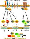
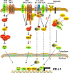
Transcription regulation network of PD-L1 in the tumor microenvironment
Fig. 6
Fig. 6
Role of non-coding RNAs in regulating PD-L1

Similar articles

Cited by

References

    1. Jiang XJ, Wang J, Deng XY, Li XL, Li XY, Zeng ZY, et al. Immunotherapy targeted to immune checkpoint: a revolutionary breakthrough in cancer therapy. Prog Biochem Biophys. 2018;45(11):1178–86.
    1. Jiang X, Shapiro DJ. The immune system and inflammation in breast cancer. Mol Cell Endocrinol. 2014;382(1):673–682. doi: 10.1016/j.mce.2013.06.003. - DOI - PMC - PubMed
    1. Wang YA, Li XL, Mo YZ, Fan CM, Tang L, Xiong F, et al. Effects of tumor metabolic microenvironment on regulatory T cells. Mol Cancer. 2018;17(1):168. doi: 10.1186/s12943-018-0913-y. - DOI - PMC - PubMed
    1. Yu J, Du W, Yan F, Wang Y, Li H, Cao S, et al. Myeloid-derived suppressor cells suppress antitumor immune responses through IDO expression and correlate with lymph node metastasis in patients with breast cancer. J Immunol. 2013;190(7):3783–3797. doi: 10.4049/jimmunol.1201449. - DOI - PubMed
    1. Benencia F, Muccioli M, Alnaeeli M. Perspectives on reprograming cancer-associated dendritic cells for anti-tumor therapies. Front Oncol. 2014;4:72. doi: 10.3389/fonc.2014.00072. - DOI - PMC - PubMed

Publication types

MeSH terms