. 2019 Apr 15;25(8):2610-2620.
doi: 10.1158/1078-0432.CCR-18-1527. Epub 2019 Jan 15.

Fucosylation Enhances the Efficacy of Adoptively Transferred Antigen-Specific Cytotoxic T Lymphocytes

Affiliations

Fucosylation Enhances the Efficacy of Adoptively Transferred Antigen-Specific Cytotoxic T Lymphocytes

Gheath Alatrash et al. Clin Cancer Res. .

Abstract

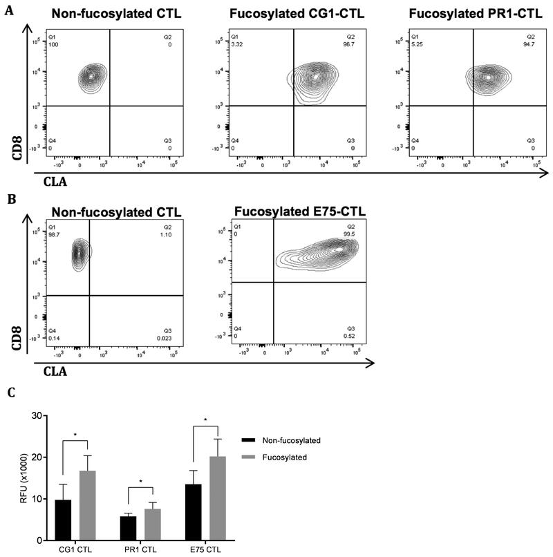
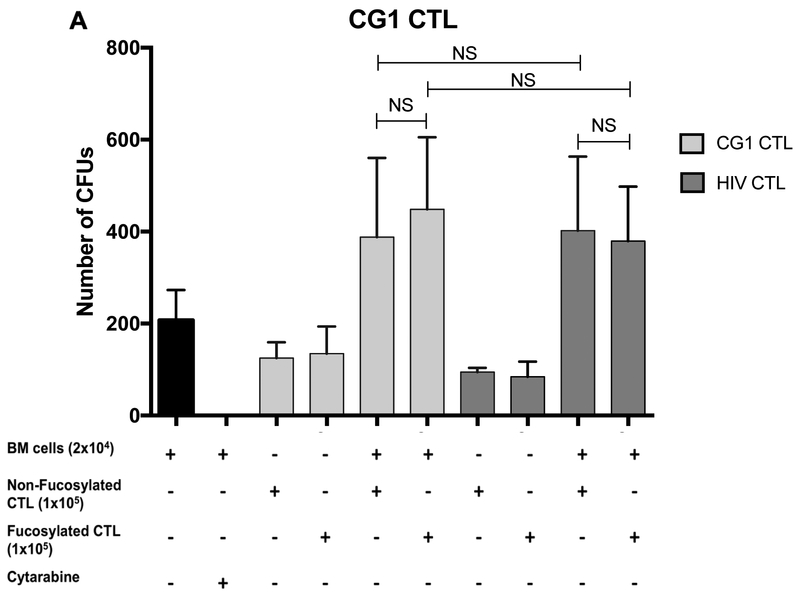
Purpose: Inefficient homing of adoptively transferred cytotoxic T lymphocytes (CTLs) to tumors is a major limitation to the efficacy of adoptive cellular therapy (ACT) for cancer. However, through fucosylation, a process whereby fucosyltransferases (FT) add fucose groups to cell surface glycoproteins, this challenge may be overcome. Endogenously fucosylated CTLs and ex vivo fucosylated cord blood stem cells and regulatory T cells were shown to preferentially home to inflamed tissues and marrow. Here, we show a novel approach to enhance CTL homing to leukemic marrow and tumor tissue.

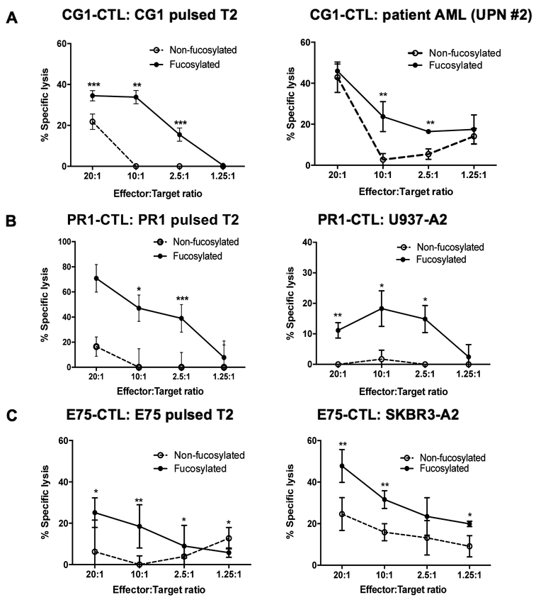
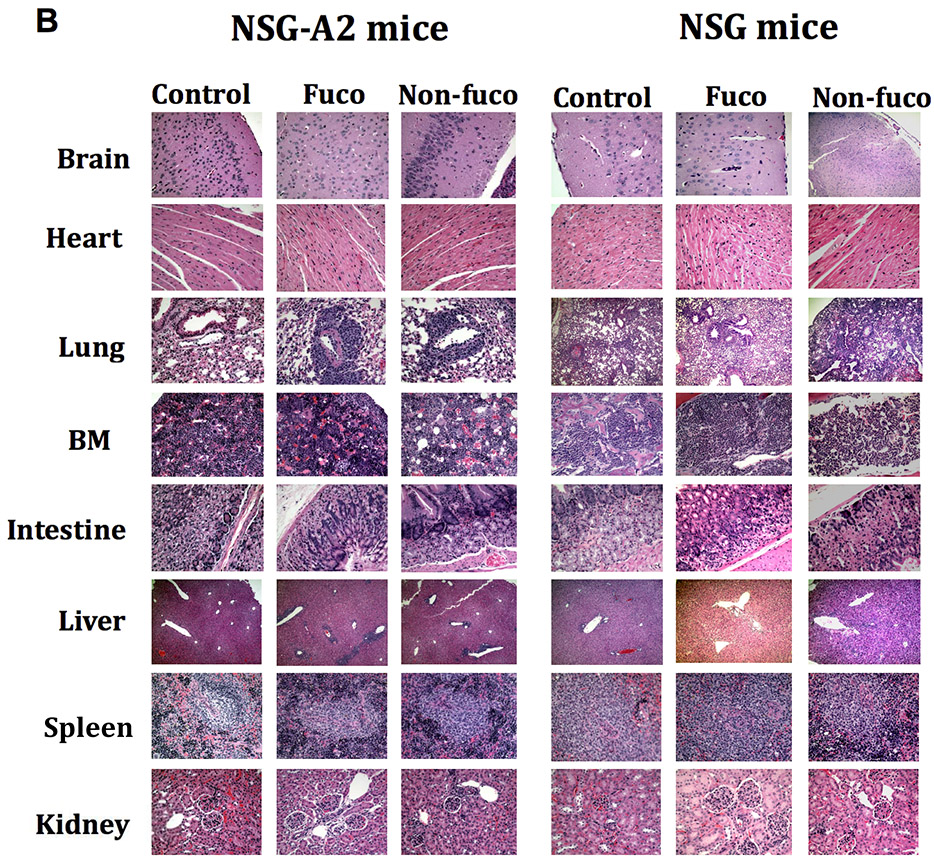
Experimental design: Using the enzyme FT-VII, we fucosylated CTLs that target the HLA-A2-restricted leukemia antigens CG1 and PR1, the HER2-derived breast cancer antigen E75, and the melanoma antigen gp-100. We performed in vitro homing assays to study the effects of fucosylation on CTL homing and target killing. We used in vivo mouse models to demonstrate the effects of ex vivo fucosylation on CTL antitumor activities against leukemia, breast cancer, and melanoma.

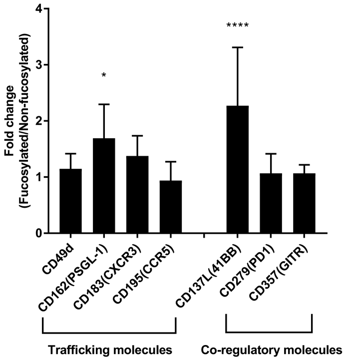
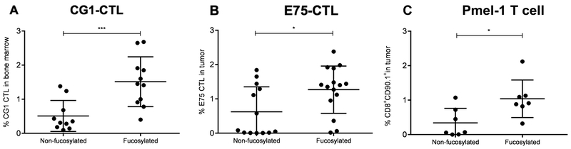
Results: Our data show that fucosylation increases in vitro homing and cytotoxicity of antigen-specific CTLs. Furthermore, fucosylation enhances in vivo CTL homing to leukemic bone marrow, breast cancer, and melanoma tissue in NOD/SCID gamma (NSG) and immunocompetent mice, ultimately boosting the antitumor activity of the antigen-specific CTLs. Importantly, our work demonstrates that fucosylation does not interfere with CTL specificity.

Conclusions: Together, our data establish ex vivo CTL fucosylation as a novel approach to improving the efficacy of ACT, which may be of great value for the future of ACT for cancer.


Conflict of interest statement

Conflict of Interest: The authors have no conflict of interest to disclose.

Figures

Figure 1.
Figure 1.. Fucosylation enhances CTL migration.
CD8 enriched antigen specific CTL were fucosylated with FT-VII. Results show representative HECA-452 staining by flow cytometry of A, leukemia specific CTL and B, breast cancer specific CTL. C, Fucosylated or non-fucosylated antigen specific CTL were passed through a HUVEC barrier to assess functional alterations to transmigration. LeukoTracker fluorescence was measured in the post-barrier well. Relative fluorescence units (RFU) are indicative of three independent experiments. Statistical testing was performed using unpaired t-test (*p < 0.05).
Figure 2.
Figure 2.. Fucosylation alters expression of select CTL surface markers.
Fucosylated antigen specific CTL were assessed for known trafficking and regulatory cell surface molecules using flow cytometry. Fucosylation of antigen-specific CTL increased the cell surface expression of a number of trafficking molecules. Cell surface expression of CD162 (PSGL-1) (1.7-fold) and CD137 (41BB) (3-fold) was significantly increased in comparison to non-fucosylated CTL. Results are representative of 6 independent experiments. Statistical testing was performed using ANOVA (*p < 0.05; ****p < 0.0001).
Figure 3.
Figure 3.. Fucosylation enhances targeted cytotoxicity in vitro.
Antigen-specific CTL were co-cultured with cognate peptide-MHC expressing target cells at varying E (effector):T (target) ratios in a calcein-AM release cytotoxicity assay. Results are depicted as % specific lysis of target cells. A, CG1-CTL and B, PR1-CTL were co-cultured with T2 cells pulsed with corresponding peptide, patient AML (UPN#2) or U937-A2 cells. Similarly, C, E75-CTL were co-cultured with E75 peptide-pulsed T2 cells or SKBR3-A2 cells at varying E:T ratios. Results are representative of 3 independent experiments. Statistics were obtained using unpaired t-test (*p < 0.05; **p < 0.005; ***p < 0.001).
Figure 4.
Figure 4.. Fucosylation enhances the efficacy of CG1-, PR1- and E75-CTL in vivo.
Irradiated NSG mice were engrafted with primary patient AML UPN#1 (1 × 10⁶ to 1 × 10⁷ cells) on day 0. Fucosylated or non-fucosylated A, CG1- or B, PR1- CTL were injected on day 2 (0.5 × 10⁶). Mice were sacrificed and bone marrow was harvested on week 2 and analyzed by flow cytometry. Results are expressed as percent hCD33+/hCD45+/mCD45 cells of total live cells in bone marrow. C, For the breast cancer model, irradiated NSG mice were engrafted with SKBR3-A2 (1.5 × 10⁷ cells) in the mammary fat pad on day 0. Fucosylated or non-fucosylated E75-CTL (2 × 10⁶ cells) were injected IV on days 7 and 10. Mice were sacrificed at week 5 and tumor sizes were measured using bioluminescence imaging. Results are expressed as percent tumor size following treatment with CTL in comparison with pre-treatment tumor size. D, C57BL/6 mice were inoculated with B16-F10 melanoma cells (0.3 × 10⁶). Fucosylated or non-fucosylated expanded pmel-1 CD8+ T cells (5 × 10⁶) were injected IV on day 0. Mice tumor volumes were measured daily for 9 days. Results are expressed as mean tumor size on day 9 from two separate experiments. Statistical testing was performed using ANOVA (*p < 0.05; **p < 0.005).
Figure 5.
Figure 5.. Fucosylation enhances CTL homing in vivo.
Irradiated tumor-bearing NSG mice treated with fucosylated or non-fucosylated CTL were analyzed for CTL homing into tumor using flow cytometry. A, Results show percent of CG1-CTL (hCD33/hCD45+/mCD45/hCD13/hCD3+/hCD8+) of total live cells in bone marrow harvested from leukemia-engrafted mice. B, Breast cancer engrafted mice were sacrificed at week 5, and TILs were enriched from the tumors. Results show percentage of E75-CTL (hCD45+/ mCD45/hCD3+/ hCD8+) of total live cells from TIL extraction. C, Melanoma-engrafted C57BL/6 mice were sacrificed on Day 9, and TILs were extracted from the primary tumor. Results show percent of pmel-1 T cells (mCD45+/mCD3+/mCD8+/CD90.1+) of total live cells. Statistical testing was performed using unpaired t-test (*p < 0.05; ***p < 0.001).
Figure 6.
Figure 6.. Fucosylation does not alter CTL specificity or homing to normal tissue.
A, Healthy donor bone marrow was co-cultured with fucosylated or non-fucosylated CG1-CTL at a 1:5 ratio of BM:CTL. Cultures were grown in Mammocult medium in a 6-well plate for 14 days before CFU counts were taken. Results show CFU on day 14 for CG1-CTL. BM co-cultured with cytarabine (1 mM) served as a positive killing control. Data is representative of 3 independent experiments. B, Fucosylated (fuco) and non-fucosylated (non-fuco) antigen-specific CTL were administered to mice twice weekly IV via tail vein. After 4 weeks, mice were sacrificed and tissues were fixed, paraffin-embedded, sectioned, and stained with hematoxylin and eosin prior to histological examination. Data show equal lymphocytic infiltration in normal mouse tissue between fuco CTL and non-fuco CTL treated groups. Statistical testing was performed using ANOVA. BM, bone marrow; NS, not significant.

References

    1. Sackstein R, Schatton T, Barthel SR. T-lymphocyte homing: an underappreciated yet critical hurdle for successful cancer immunotherapy. Lab Invest 2017;97(6):669–97 doi 10.1038/labinvest.2017.25. - DOI - PMC - PubMed
    1. Abastado JP. The next challenge in cancer immunotherapy: controlling T-cell traffic to the tumor. Cancer Res 2012;72(9):2159–61 doi 10.1158/0008-5472.CAN-11-3538. - DOI - PubMed
    1. Ley K, Laudanna C, Cybulsky MI, Nourshargh S. Getting to the site of inflammation: the leukocyte adhesion cascade updated. Nature reviews Immunology 2007;7(9):678–89 doi 10.1038/nri2156. - DOI - PubMed
    1. Kurachi M, Kurachi J, Suenaga F, Tsukui T, Abe J, Ueha S, et al. Chemokine receptor CXCR3 facilitates CD8(+) T cell differentiation into short-lived effector cells leading to memory degeneration. The Journal of experimental medicine 2011;208(8):1605–20 doi 10.1084/jem.20102101. - DOI - PMC - PubMed
    1. Bromley SK, Mempel TR, Luster AD. Orchestrating the orchestrators: chemokines in control of T cell traffic. Nat Immunol 2008;9(9):970–80 doi 10.1038/ni.f.213. - DOI - PubMed

Publication types