. 2019 Jan 29;116(5):1698-1703.
doi: 10.1073/pnas.1803999116. Epub 2019 Jan 15.

Suppression of chemotherapy-induced cytokine/lipid mediator surge and ovarian cancer by a dual COX-2/sEH inhibitor

Affiliations

Suppression of chemotherapy-induced cytokine/lipid mediator surge and ovarian cancer by a dual COX-2/sEH inhibitor

Allison Gartung et al. Proc Natl Acad Sci U S A. .

Abstract

Although chemotherapy is a conventional cancer treatment, it may induce a protumorigenic microenvironment by triggering the release of proinflammatory mediators. In this study, we demonstrate that ovarian tumor cell debris generated by first-line platinum- and taxane-based chemotherapy accelerates tumor progression by stimulating a macrophage-derived "surge" of proinflammatory cytokines and bioactive lipids. Thus, targeting a single inflammatory mediator or pathway is unlikely to prevent therapy-induced tumor progression. Here, we show that combined pharmacological abrogation of the cyclooxygenase-2 (COX-2) and soluble epoxide hydrolase (sEH) pathways prevented the debris-induced surge of both cytokines and lipid mediators by macrophages. In animal models, the dual COX-2/sEH inhibitor PTUPB delayed the onset of debris-stimulated ovarian tumor growth and ascites leading to sustained survival over 120 days postinjection. Therefore, dual inhibition of COX-2/sEH may be an approach to suppress debris-stimulated ovarian tumor growth by preventing the therapy-induced surge of cytokines and lipid mediators.

Keywords: cyclooxygenase; debris; inflammation; oxylipins; soluble epoxide hydrolase.


Conflict of interest statement

Conflict of interest statement: M.W.K. is now an employee of Bristol-Myers Squibb. His position at Bristol-Myers Squibb is not related to this work.

Figures

Fig. 1.
Fig. 1.
Chemotherapy-generated ovarian tumor cell debris stimulates tumor growth and shortens survival. Percent survival of mice coinjected intraperitoneally with (A) cisplatin- or (B) paclitaxel-generated ID8 debris (9 × 10⁵ dead cells) and ID8 living cells (1 × 10⁶). n = 5 mice per group. Kaplan–Meier analysis indicated significantly shortened survival in mice coinjected with (A) cisplatin- (log-rank test = 9.65, *P = 0.0019) or (B) paclitaxel- (log-rank test = 9.85, *P = 0.0017) generated ID8 debris and ID8 living cells compared with ID8 living cells alone. Image shows representative mice on day 56 postinjection. Yellow dashed circle indicates ascites in mice coinjected with paclitaxel-generated debris and ID8 living cells. (C) Percent survival of mice coinjected orthotopically with paclitaxel-generated ID8 debris (9 × 10⁵ dead cells) and ID8 living cells (1 × 10⁶). n = 5 mice per group. Kaplan–Meier analysis indicated significantly shortened survival in mice coinjected with paclitaxel-generated ID8 debris and ID8 living cells compared with ID8 living cells alone (log-rank test = 6.00, *P = 0.014).
Fig. 2.
Fig. 2.
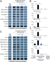
Cytokine surge by debris-stimulated macrophages is prevented by the dual COX-2/sEH inhibitor PTUPB. (A) Cytokine array of conditioned medium from RAW264.7 murine macrophages treated with vehicle or PTUPB and stimulated with paclitaxel-generated ID8 debris. (B) ELISA quantification of proinflammatory cytokines released by RAW264.7 macrophages treated with vehicle (black bars) or PTUPB (5 μM) (blue bars) for 2 h and stimulated with paclitaxel-generated ID8 debris, or by paclitaxel-generated ID8 debris alone without macrophages. Data are presented as means (pg/mL) ± SEM n = 7–8/group. *P < 0.05 vs. RAW264.7 + paclitaxel-generated ID8 dead cells. n.d., not detectable. (C) Cytokine array of conditioned medium from RAW264.7 murine macrophages treated with vehicle or PTUPB for 2 h and stimulated with carboplatin-generated ID8 debris.
Fig. 3.
Fig. 3.
Dual COX-2/sEH inhibition differentially regulates the release of lipid mediators by debris-stimulated macrophages. LC-MS/MS–based oxylipin analysis of conditioned medium from paclitaxel-generated ID8 debris alone without macrophages (“debris alone,” blue bars) or from RAW264.7 macrophages stimulated with paclitaxel-generated ID8 debris (orange bars), PTUPB-treated macrophages (5 μM, 2 h) stimulated with paclitaxel-generated ID8 debris (gray bars), or macrophages not stimulated with debris (“macrophages alone,” yellow bars). PTUPB inhibits the surge of PGF, PGD2, and PGJ2 (Left), while neutralizing the reduction of PGE2, THF diol, 15-HETE, 11-HETE, and 5-HETE (Center) by debris-stimulated macrophages. PTUPB suppressed the release of 15-oxoETE by macrophages (Right). Data are presented as means (pmol/L) ± SEM n = 10 per group. *P < 0.05 or **P < 0.01 vs. macrophages + debris or macrophages alone.
Fig. 4.
Fig. 4.
PTUPB suppresses proangiogenic cytokines in vivo. Proangiogenic cytokine arrays of (A) serum or (B) ascites from control or PTUPB-treated mice intraperitoneally injected with ID8 (1 × 10⁶ living cells). Serum and ascites was collected on day 60 postinjection.
Fig. 5.
Fig. 5.
Suppression of debris-stimulated ovarian tumor growth by PTUPB. Percent survival of mice coinjected intraperitoneally with (A) paclitaxel- or (B) carboplatin-generated ID8 debris (9 × 10⁵ dead cells) and ID8 living cells (1 × 10⁶), or (C) coinjected orthotopically with paclitaxel-generated ID8 debris (9 × 10⁵ dead cells) and ID8 living cells (1 × 10⁶). Image shows representative orthotopic tumors on day 62 postinjection. (Scale bar,1 cm.) Systemic treatment with PTUPB (30 mg/kg/d) or control initiated 4 wk postinjection. n = 4–5 mice per group. Kaplan–Meier analysis and log-rank testing indicated significantly prolonged survival in mice treated with PTUPB compared with control in the (A) paclitaxel-generated (log-rank test = 9.70, *P = 0.0018) and (B) carboplatin-generated (log-rank test = 9.85, *P = 0.0017) debris-stimulated intraperitoneal ovarian tumor models, as well as the (C) paclitaxel-generated debris-stimulated orthotopic ovarian tumor model (log-rank test = 7.91, *P = 0.005).

References

    1. Thibault B, Castells M, Delord JP, Couderc B. Ovarian cancer microenvironment: Implications for cancer dissemination and chemoresistance acquisition. Cancer Metastasis Rev. 2014;33:17–39. - PubMed
    1. Sulciner ML, et al. Resolvins suppress tumor growth and enhance cancer therapy. J Exp Med. 2018;215:115–140. - PMC - PubMed
    1. Chang J, et al. Chemotherapy-generated cell debris stimulates colon carcinoma tumor growth via osteopontin. FASEB J. 2018 doi: 10.1096/fj.201800019RR. - DOI - PMC - PubMed
    1. Revesz L. Effect of tumour cells killed by X-rays upon the growth of admixed viable cells. Nature. 1956;178:1391–1392. - PubMed
    1. Huang Q, et al. Caspase 3-mediated stimulation of tumor cell repopulation during cancer radiotherapy. Nat Med. 2011;17:860–866. - PMC - PubMed

Publication types

MeSH terms