. 2019 Jan 29;116(5):1755-1764.
doi: 10.1073/pnas.1816933116. Epub 2019 Jan 15.

Human cytomegalovirus G protein-coupled receptor US28 promotes latency by attenuating c-fos

Affiliations

Human cytomegalovirus G protein-coupled receptor US28 promotes latency by attenuating c-fos

Benjamin A Krishna et al. Proc Natl Acad Sci U S A. .

Abstract

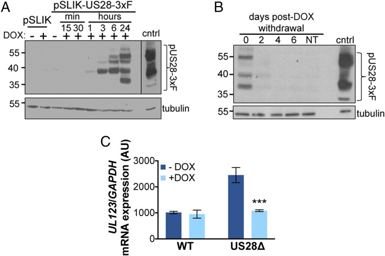
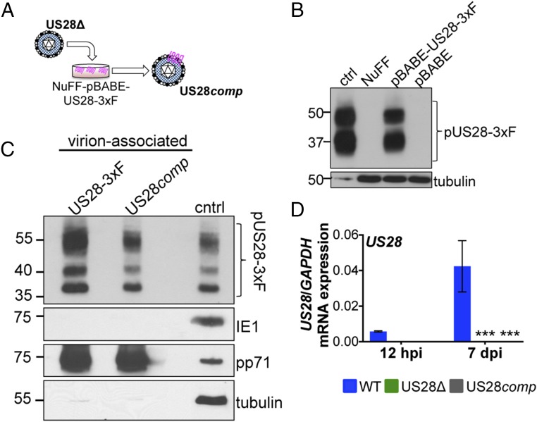
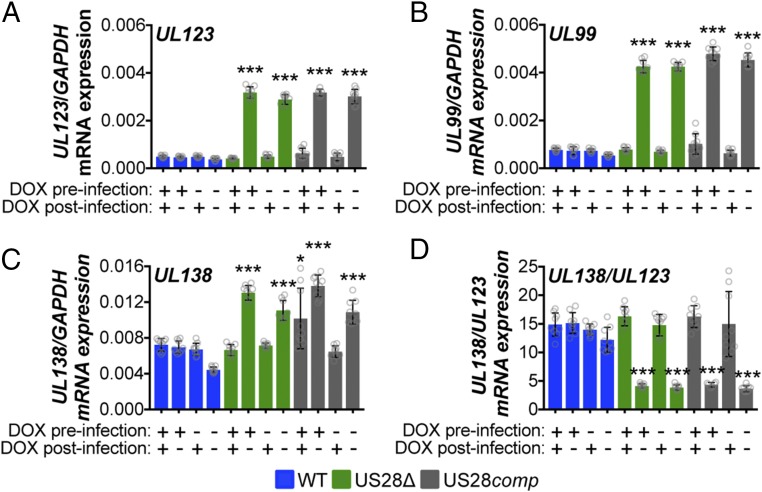
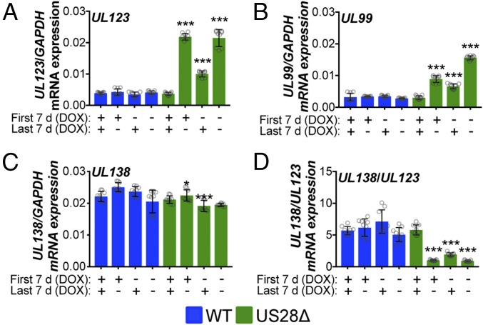
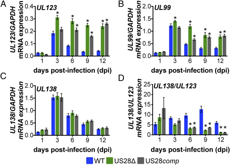
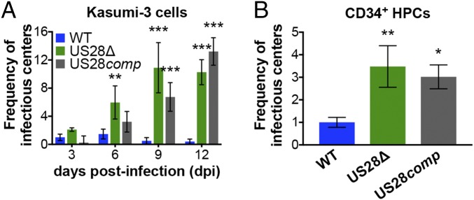
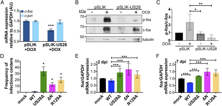
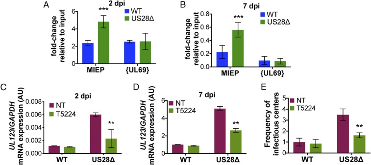
Human cytomegalovirus (HCMV) is a ubiquitous pathogen that undergoes latency in cells of the hematopoietic compartment, although the mechanisms underlying establishment and maintenance of latency remain elusive. We previously reported that the HCMV-encoded G protein-coupled receptor (GPCR) homolog US28 is required for successful latent infection. We now show that US28 protein (pUS28) provided in trans complements the US28Δ lytic phenotype in myeloid cells, suggesting that sustained US28 expression is necessary for long-term latency. Furthermore, expression of pUS28 at the time of infection represses transcription from the major immediate early promoter (MIEP) within 24 h. However, this repression is only maintained in the presence of continual pUS28 expression provided in trans Our data also reveal that pUS28-mediated signaling attenuates both expression and phosphorylation of cellular fos (c-fos), an AP-1 transcription factor subunit, to repress MIEP-driven transcription. AP-1 binds to the MIEP and promotes lytic replication, and in line with this we find that US28Δ infection results in an increase in AP-1 binding to the MIEP, compared with WT latent infection. Pharmacological inhibition of c-fos represses the MIEP during US28Δ infection to levels similar to those we observe during WT latent infection. Together, our data reveal that US28 is required for both establishment and long-term maintenance of HCMV latency, which is modulated, at least in part, by repressing functional AP-1 binding to the MIEP.

Keywords: HCMV; US28; cytomegalovirus; herpesvirus; latency.


Conflict of interest statement

The authors declare no conflict of interest.

Figures

Fig. 1.
Fig. 1.
Continual pUS28 expression complements the lytic-like phenotype following US28Δ infection. (A) THP-1 cells were transduced with THP-1-pSLIK-hygro (pSLIK) or THP-1-pSLIK-US28-3xF (pSLIK-US28-3xF) and treated with (+) or without (−) DOX (1 μg/mL) to induce pUS28 expression. Cells were harvested at the indicated time points posttreatment. pUS28 expression was detected by immunoblot using the FLAG epitope tag. (B) THP-1-pSLIK-US28-3xF cells were treated with DOX for 24 h and harvested to confirm pUS28 expression (0 d). Remaining cells were washed in PBS and cultured in the absence of DOX for the duration of the experiment. Cell samples were taken at the indicated days posttreatment and all samples were then immunoblotted for pUS28 using the epitope tag. (A and B) As a control (cntrl), NuFF-1 cells were infected with US28-3xF (moi = 0.5) and cell lysates were harvested at 96 h postinfection. Tubulin is shown as a loading control. Note that due to the intensity of this control lysate in A, a shorter exposure is shown, as denoted by the black line. (C) THP-1-pSLIK-US28-3xF cells were treated without (−; dark blue) or with (+; light blue) DOX for 24 h and then infected with WT or US28Δ (moi = 1.0). DOX was replenished every 48 h and the cells were harvested at 6 dpi for UL123 expression by RT-qPCR. Samples were normalized to GAPDH, and each sample was analyzed in triplicate. Errors bars indicate SD. Statistical significance was calculated using Welch’s t test; ***P < 0.001.
Fig. 2.
Fig. 2.
US28comp incorporates pUS28 into its virion but fails to express US28 after infection. (A) US28Δ was grown on NuFF-pBABE-US28-3xF cells. Cell-free virus was isolated and termed US28comp. (B) Lysates from NuFF-1 cells infected with TB40/E-mCherry-US28-3xF (US28-3xF; moi = 0.5, 96 h postinfection, 15 μg; ctrl), mock-infected NuFF-1 cells (30 μg; NuFF), or NuFF-1 cells stably transduced with either pBABE-US28-3xF (30 μg; NuFF-pBABE-US28-3xF) or pBABE (30 μg; NuFF-pBABE) were assessed for pUS28 expression by immunoblot using an antibody directed at the FLAG epitope tag. Tubulin is shown as a loading control. (C) US28comp virus was generated by infecting NuFF-pBABE-US28-3xF with US28Δ. As a control for virion-associated pUS28, NuFF-1 cells were infected with US28-3xF. Cell-free US28comp or US28-3xF virus was purified through a sorbitol cushion and pUS28 was detected by immunoblot for the FLAG epitope tag. Cell lysates (15 μg) from NuFF-1 cells infected with US28-3xF (moi = 0.5, 96 hpi) are shown as a control (cntrl). Samples were also probed with antibodies directed at the viral proteins IE1 and pp71, as well as cellular tubulin. (D) Kasumi-3 cells were infected with WT, US28Δ, or US28comp at a multiplicity of 1.0 TCID50 per cell and harvested 7 dpi. US28 gene expression was quantified by RT-qPCR using primers that amplify US28 (SI Appendix, Table S1). Samples were normalized to GAPDH, and each sample was analyzed in triplicate. Errors bars indicate SD. Statistical significance was calculated using two-way ANOVA and Dunnett’s post hoc analysis relative to WT at each time point; ***P < 0.001.
Fig. 3.
Fig. 3.
Virion-delivered pUS28 fails to maintain suppression of lytic gene transcription. THP-1-pSLIK-US28-3xF were treated with (+) or without (−) DOX (1 μg/mL) to induce pUS28 expression (preinfection) and then infected with WT (blue), US28Δ (green), or US28comp (gray) (moi = 1.0). Infected cells were washed 2 h postinfection and cultured in the presence (+) or absence (−) of DOX, as indicated (postinfection), where cultures receiving postinfection DOX were replenished every 48 h. Cells were harvested 12 dpi and (A) UL123, (B) UL99, (C) UL138, and (D) the ratio of UL138/UL123 expression were measured by RT-qPCR. Samples were normalized to GAPDH and analyzed in triplicate. Errors bars indicate SD and statistical significance was calculated using one-way ANOVA and Tukey post hoc analysis; *P < 0.05, ***P < 0.001.
Fig. 4.
Fig. 4.
Sustained pUS28 expression suppresses lytic gene expression. THP-1-pSLIK-US28-3xF cells were treated with (+) or without (−) DOX (1 μg/mL) for 48 h to induce pUS28 expression and then infected with WT (blue) or US28Δ (green) (moi = 1.0). Cells were cultured for 7 d, during which cells were maintained under their original treatment conditions (First 7 d). At 7 dpi, cells were washed and treated with (+) or without (−) DOX (Last 7 d). Total RNA was harvested and (A) UL123, (B) UL99, (C) UL138, and (D) the ratio of UL138/UL123 expression were measured by RT-qPCR. Samples were normalized to GAPDH and assessed in triplicate. Errors bars indicate SD and significance calculated using one-way ANOVA and Dunnett’s post hoc analysis; *P < 0.05, ***P < 0.001.
Fig. 5.
Fig. 5.
pUS28 represses UL123 and UL99 expression in infected Kasumi-3 cells at early times of latent infection. Kasumi-3 cells were infected with WT (blue), US28Δ (green), or US28comp (gray) (moi = 1.0) and sorted for mCherry-positive cells at 1 dpi. Cells were then harvested at the indicated dpi. (A) UL123, (B) UL99, (C) UL138, and (D) the ratio of UL138/UL123 expression were measured by RT-qPCR. Samples were normalized to GAPDH and analyzed in triplicate. Errors bars indicate SD, and statistical significance was calculated using two-way ANOVA and Dunnett’s post hoc analysis relative to WT virus at each time point; *P < 0.05.
Fig. 6.
Fig. 6.
Sustained pUS28 expression is required to maintain viral latency. (A) Kasumi-3 cells (moi = 1.0) or (B) CD34+ HPCs (moi = 2.0) were infected with WT (blue), US28Δ (green), or US28comp (gray). (A and B) Cells were collected at the indicated time points and reactivation events were determined by ELDA. Data in A are presented as fold change in virus release relative to WT virus on day 3. Data in B are presented as fold change in virus release relative to WT virus. In all cases, error bars represent SDs of three biological replicates. Statistical significance was calculated relative to WT at the same time point using (A) two-way ANOVA analysis or (B) one-way ANOVA and Tukey post hoc analysis; *P < 0.05, **P < 0.01, ***P < 0.001.
Fig. 7.
Fig. 7.
US28 signaling attenuates both c-fos expression and activation. (A) THP-1-pSLIK-hygro (pSLIK) and THP-1-pSLIK-US28-3xF (pSLIK-US28) were treated with DOX and cells were harvested 24 h posttreatment. c-fos (dark blue) and c-jun (light blue) expression were measured relative to GAPDH. (B) Cells from A were treated with (+) or without (−) DOX and cells were harvested 24 h posttreatment. Phosphorylated fos (p-fos), total fos (c-fos), and tubulin were detected by immunoblot. Representative immunoblot shown (n = 3). (C) Quantification of the results shown in B using densitometry. Levels of p-fos were quantified relative to c-fos, shown relative to tubulin. Data were quantified from three biological replicates. Error bars indicate SD. Statistical significance was calculated using one-way ANOVA and Tukey post hoc analysis. (DF) Kasumi-3 cells were infected with the indicated viruses (moi = 1.0) and harvested at (E) 2 or (D and F) 7 dpi. (D) The frequency of infectious virus from each latently infected culture was determined by ELDA. Data are presented as fold change in virus release relative to WT. (E and F) c-fos expression was measured relative to GAPDH. (A and DF) Each sample was analyzed in triplicate. Error bars indicate SD. Statistical significance was calculated using Welch’s t test; *P < 0.05, **P < 0.01, ***P < 0.001.
Fig. 8.
Fig. 8.
pUS28 decreases c-fos binding at the MIEP, leading to transcriptional repression of IE transcripts. (A and B) Kasumi-3 cells were infected (moi = 1.0) with WT (blue) or US28Δ (green). Cells were collected (A) 2 or (B) 7 dpi and the AP-1 complex was immunoprecipitated using an anti–c-fos antibody. Coprecipitated MIEP was quantified by qPCR, and data are shown as fold change relative to input. The UL69 nonpromoter region is shown as a control. (CE) Kasumi-3 cells were infected as in A and B in the absence (red; NT) or presence (green) of the fos inhibitor, T5224 (10 nM), and cells were harvested (C) 2 or (D and E) 7 dpi. (C and D) UL123 expression was measured and normalized to GAPDH. (E) The frequency of infectious virus from each latently infected culture was determined by ELDA. Data are presented as fold change in virus release relative to vehicle-treated WT. Each sample was analyzed in triplicate. Errors bars indicate SD and statistical significance was measured using Welch’s t test; **P < 0.01, ***P < 0.001.

Similar articles

Cited by

References

    1. Mocarski ES, Courcelle CT. Cytomegaloviruses and Their Replication. 4th Ed Lippincott Williams & Wilkins; Philadelphia: 2001.
    1. Pass RF. Cytomegaloviruses. 4th Ed Lippincott Williams & Wilkins; Philadelphia: 2001.
    1. Khanna R, Diamond DJ. Human cytomegalovirus vaccine: Time to look for alternative options. Trends Mol Med. 2006;12:26–33. - PubMed
    1. Arvin AM, Fast P, Myers M, Plotkin S, Rabinovich R. National Vaccine Advisory Committee Vaccine development to prevent cytomegalovirus disease: Report from the National Vaccine Advisory Committee. Clin Infect Dis. 2004;39:233–239. - PubMed
    1. Griffiths P, Baraniak I, Reeves M. The pathogenesis of human cytomegalovirus. J Pathol. 2015;235:288–297. - PubMed

Publication types

MeSH terms